. 2009 May 27;4(5):e5702.
doi: 10.1371/journal.pone.0005702.

CD152 (CTLA-4) determines CD4 T cell migration in vitro and in vivo

Affiliations

CD152 (CTLA-4) determines CD4 T cell migration in vitro and in vivo

Karin Knieke et al. PLoS One. .

Abstract

Background: Migration of antigen-experienced T cells to secondary lymphoid organs and the site of antigenic-challenge is a mandatory prerequisite for the precise functioning of adaptive immune responses. The surface molecule CD152 (CTLA-4) is mostly considered as a negative regulator of T cell activation during immune responses. It is currently unknown whether CD152 can also influence chemokine-driven T cell migration.

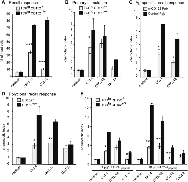
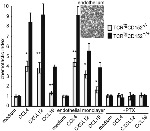
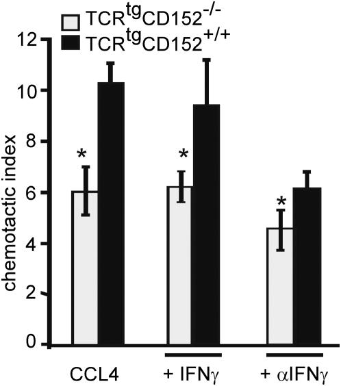
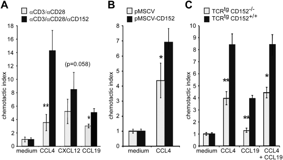
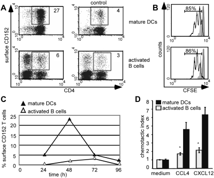
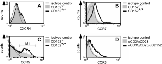
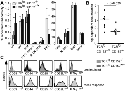
Methodology/principal findings: We analyzed the consequences of CD152 signaling on Th cell migration using chemotaxis assays in vitro and radioactive cell tracking in vivo. We show here that the genetic and serological inactivation of CD152 in Th1 cells reduced migration towards CCL4, CXCL12 and CCL19, but not CXCL9, in a G-protein dependent manner. In addition, retroviral transduction of CD152 cDNA into CD152 negative cells restored Th1 cell migration. Crosslinking of CD152 together with CD3 and CD28 stimulation on activated Th1 cells increased expression of the chemokine receptors CCR5 and CCR7, which in turn enhanced cell migration. Using sensitive liposome technology, we show that mature dendritic cells but not activated B cells were potent at inducing surface CD152 expression and the CD152-mediated migration-enhancing signals. Importantly, migration of CD152 positive Th1 lymphocytes in in vivo experiments increased more than 200% as compared to CD152 negative counterparts showing that indeed CD152 orchestrates specific migration of selected Th1 cells to sites of inflammation and antigenic challenge in vivo.

Conclusions/significance: We show here, that CD152 signaling does not just silence cells, but selects individual ones for migration. This novel activity of CD152 adds to the already significant role of CD152 in controlling peripheral immune responses by allowing T cells to localize correctly during infection. It also suggests that interference with CD152 signaling provides a tool for altering the cellular composition at sites of inflammation and antigenic challenge.


Conflict of interest statement

Competing Interests: The authors have declared that no competing interests exist.

Figures

Figure 1
Figure 1. CD152 enhances chemotaxis of Th1 cells.
(A) Migration of unpolarized T cells. Recall response of CD4+ OVA-specific TCRtg T cells from CD152−/− or CD152+/+ mice was induced by adding 1 µg/ml OVA323–339 and T cell-depleted APCs. On day 6 of recall response cells were analyzed in chemotaxis assays. Bars indicate migrated CD4+ cells as percentage of input cells. (B) Migration of CD152−/− and CD152+/+ T cells in primary stimulation: CD4+ OVA-specific TCRtg T cells were stimulated with 1 µg/ml OVA323–339 and T cell-depleted APCs. On day 6 of primary stimulation cells were analyzed in chemotaxis assays. Bars show the chemotactic index of CD4+ cells. (C) Specific migration of antigen-specific stimulated Th1 cells in a recall response: Primary stimulation and recall response of CD4+CD62L+ OVA-specific TCRtg T cells were performed under Th1 conditions with 1 µg/ml OVA-peptide in the presence of 200 µg/ml neutralizing anti-CD152 Fab fragments or hamster control Fab fragments. Cells were examined in chemotaxis assay on day 6 of recall response. (D) Chemotactic index of a polyclonally induced recall response of CD152−/− or CD152+/+ Th1 cells: Primary stimulation and recall response of splenocytes from CD152−/− and CD152+/+ mice were induced polyclonally (as described in Material and Methods) and CD4+ cells were used for chemotaxis assays on day 4 of recall response. (E) Migration of CD152−/− and CD152+/+ Th1 cells in a recall response is dose dependent: Primary stimulation and recall response of CD4+ cells from TCRtg CD152−/− and CD152+/+ mice were induced antigen-specifically using Th1 conditions with indicated amounts of OVA-peptide and cells were analyzed on day 6 of recall response in chemotaxis assays. All data shown represent one out of 3–4 similar experiments.
Figure 2
Figure 2. Only CD152+/+ but not CD152−/− cells show efficient transendothelial migration.
Primary stimulation and recall response of CD4+ OVA-specific TCRtg T cells from CD152−/− and CD152+/+ mice were performed using Th1 conditions with 10 µg/ml OVA-peptide and T cell-depleted APCs. On day 6 of recall response migration of CD4+ cells through membranes coated with or without endothelial monolayer was analyzed after 90 min. incubation at 37°C in Transwell chemotaxis assay. CD152 mediated migration of Th1 cells is G-Protein dependent: CD4+ OVA-specific TCRtg CD152−/− and CD152+/+ Th1 cells were incubated for 2 hours at 37°C in the presence of 100 ng/ml Pertussis toxin prior to examination in chemotaxis assay. (Inset) Endothelial cells (mlEND) were grown to a confluent monolayer for 48 h on Transwell membranes and confluency was controlled by microscopy. Shown data represent one out of 2 similar experiments.
Figure 3
Figure 3. CD152-enhanced migration is IFNγ independent.
CD4+ OVA-specific TCRtg splenocytes from CD152−/− and CD152+/+ were stimulated under Th1 conditions (as described in Fig. 2) in the presence of 20 ng/ml recombinant IFN-γ or 10 µg/ml blocking anti-IFN-γ -antibodies. On day 6 after inducing a recall response cells were analyzed for migration towards CCL4 in chemotaxis assays. The data represent one out of 3 similar experiments.
Figure 4
Figure 4. CD152-signaling mediates migration of recently activated Th1 cells.
(A) CD4+CD62L+ TCRtg T cells were stimulated with 1 µg/ml OVA peptide and T cell-depleted APCs under Th1 conditions. Recall response of Th1 cells was induced by platebound anti-CD3/anti-CD28 for 48 hours. Cells were washed, incubated with microspheres coated with anti-CD3, anti-CD28 and anti-CD152 (0.15 µg/ml, 0.4 µg/ml, 4.5 µg/ml respectively) or anti-CD3, anti-CD28 and hamIgG for additional 24 hours and examined in chemotaxis assay. (B) Restored specific migration of CD152−/− Th1 cells by CD152 expression. CD152−/− splenocytes were activated with 2 µg/ml anti-CD3 under Th1 conditions and on day 2 were retrovirally transduced with cDNA of full length CD152 (pMSCV-CD152) or with empty vector pMSCV. Chemotactic capacity of transduced CD4+ cells was determined in chemotaxis assays. (C) Migration towards CCL19 plus CCL4. CD4+ TCRtg T cells from CD152+/+ and CD152−/− mice were stimulated under Th1 conditions as described in figure 2. On day 6 of recall response viable cells were analyzed in chemotaxis assays for migration capacity towards CCL4 and CCL19 alone or in combination. One out of at least 3 experiments with similar results is shown.
Figure 5
Figure 5. CD152-enhanced migration is primarily mediated by DCs.
(A) Surface expression of CD152 on CD4+ cells is up-regulated by stimulation with DCs. Naϊve CD4+CD62L+ TCRtg T cells were stimulated with 1 µg/ml OVA-peptide presented by matured DCs or by LPS-activated B cells. After 48 hours surface expression of CD152 was detected by liposome staining technique and subsequent FACS analysis. Left panels show CD152 staining, right panels show blocking controls. Numbers indicate the percentage of CD152+ CD4+ based on total CD4+ cells. (B) Equal proliferation of T cells stimulated with activated B cells or matured DCs. Naϊve CD4+CD62L+ TCRtg T cells were labeled with CFSE and antigen-specifically stimulated with activated B cells or matured DCs. Proliferation of T cells was determined by flow cytometry 48 hours (filled curve) and 72 hours later (black line). T cells cultured with different APCs but without antigenic stimulation are shown as heavy grey line. Numbers indicate the percentage of divided T cells 72 hours after stimulation relating to unstimulated controls (after 48 h 48% of Dc-stimulated T cells and 46% of Bc-stimulated T cells proliferated). (C) Kinetics of CD152 surface expression on CD4+ T cells. Indicated time after onset of stimulation cells cultured as described in 5A were stained with liposome staining technique for CD152. Percentages of surface expressing CD152 cells of total CD4+ cells are shown. (D) Chemotactic index of Th1 cells stimulated with different APCs. CD4+CD62L+ TCRtg T cells were stimulated under Th1 conditions with 1 µg/ml OVA peptide presented by activated B-cells or bone marrow derived DCs. On day 6 of primary stimulation, CD4+ cells were analyzed for migration capacity in chemotaxis assay. All data show representative data from at least two experiments.
Figure 6
Figure 6. CD152 mediates up-regulation of chemokine receptors CCR5 and CCR7.
Recall response of splenocytes from CD152+/+ and CD152−/− mice was induced polyclonally under Th1 conditions. On day 3 of recall response, cells were stained for surface markers and analyzed by flow cytometry. The histograms indicate surface expression on CD4+ lymphocytes of (A) CXCR4, (B) CCR7 and (C) CCR5; (M1: 48% CD152+/+; 29% CD152−/−). (D) After performing recall response of splenocytes from CD152+/+ mice with microspheres (as described in Fig. 4A) CCR5 was detected on CD4+ cells. Representative results from one out of 3–5 experiments are shown.
Figure 7
Figure 7. Th1 cell homing to the site of inflammation and lymph nodes is enhanced by CD152.
(A) Distribution of TCRtg CD152−/− and CD152+/+ Th1 cells 48 hours after transfer. In vitro induced Th1 cells from a recall response were radioactively labeled and injected i.v. (5×10⁶ cells per mouse). After 24 hours OVA-peptide (250 ng) in IFA was administered s.c. into one footpad (DTH); PBS/IFA-injection in the other footpad served as control. Different organs were analyzed after 48 hours for radioactivity. Recovered radioactivity in organs was calculated as percentage of total amount of radioactivity per mouse (7 mice per group). (LN: mesenteric and axillary lymph nodes; drLN: draining (popliteal and inguinal) lymph nodes; PBL: peripheral blood lymphocytes). Results from one out of two similar experiments are shown. (B) Specific migration of transferred CD152−/− and CD152+/+ Th1 cells to the site of inflammation. Radioactively labeled CD152−/− and CD152+/+ Th1 cells were transferred into mice and DTH was induced in footpads as described in 7A. Shown is the antigen-dependent accumulation of Th1 cells to the site of inflammation indicating the ratio of recovered radioactivity in the inflamed footpad versus the PBS injected footpad. (C) Similar expression of activation induced markers and similar amount of IFN-γ-producers. TCRtg CD152−/− and CD152+/+ CD4+ T cells were either left unstimulated or were in vitro stimulated under Th1 conditions and a recall response was performed. Unstimulated cells were stained on day 3 (upper panel) and stimulated cells on day 5 of recall response for activation induced molecules (lower panel). Intracellular staining for IFN-γ of fixed cells was performed on day 3 of recall response after restimulation with PMA/Ionomycin for 6 hours; Brefeldin A was added for the last 2 hours of incubation. One out of two similar experiments is shown.

References

    1. Campbell DJ, Kim CH, Butcher EC. Chemokines in the systemic organization of immunity. Immunological Reviews. 2003;195:58–71. - PubMed
    1. Hamann A, Engelhardt B. Leukocyte trafficking. Weinheim: Wiley-VCH; 2005. p. 517.
    1. Szabo SJ, Sullivan BM, Peng SL, Glimcher LH. Molecular mechanisms regulating Th1 immune responses. Annual Review of Immunology. 2003;21:713–758. - PubMed
    1. Olsson T. Critical Influences of the Cytokine Orchestration on the Outcome of Myelin Antigen-Specific T-Cell Autoimmunity in Experimental Autoimmune Encephalomyelitis and Multiple-Sclerosis. Immunological Reviews. 1995;144:245–268. - PubMed
    1. Casares D, Brumeanu TD. Insights into the pathogeneses of type I diabetes: a hint for novel immunospecific therapies. Curr Mol Med. 2001;1:357–378. - PubMed

Publication types

MeSH terms