

Highly active microbial phosphoantigen induces rapid yet sustained MEK/Erk- and PI-3K/Akt-mediated signal transduction in anti-tumor human gammadelta T-cells
- PMID: 19479075
- PMCID: PMC2682580
- DOI: 10.1371/journal.pone.0005657
Highly active microbial phosphoantigen induces rapid yet sustained MEK/Erk- and PI-3K/Akt-mediated signal transduction in anti-tumor human gammadelta T-cells
Abstract
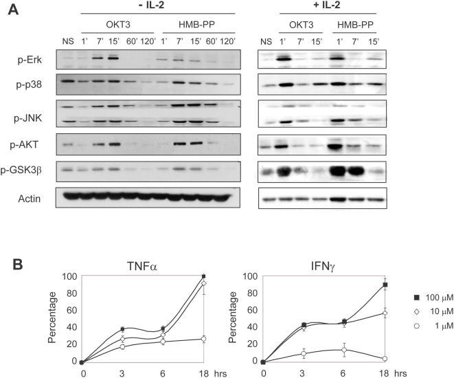
Background: The unique responsiveness of Vgamma9Vdelta2 T-cells, the major gammadelta subset of human peripheral blood, to non-peptidic prenyl pyrophosphate antigens constitutes the basis of current gammadelta T-cell-based cancer immunotherapy strategies. However, the molecular mechanisms responsible for phosphoantigen-mediated activation of human gammadelta T-cells remain unclear. In particular, previous reports have described a very slow kinetics of activation of T-cell receptor (TCR)-associated signal transduction pathways by isopentenyl pyrophosphate and bromohydrin pyrophosphate, seemingly incompatible with direct binding of these antigens to the Vgamma9Vdelta2 TCR. Here we have studied the most potent natural phosphoantigen yet identified, (E)-4-hydroxy-3-methyl-but-2-enyl pyrophosphate (HMB-PP), produced by Eubacteria and Protozoa, and examined its gammadelta T-cell activation and anti-tumor properties.
Methodology/principal findings: We have performed a comparative study between HMB-PP and the anti-CD3epsilon monoclonal antibody OKT3, used as a reference inducer of bona fide TCR signaling, and followed multiple cellular and molecular gammadelta T-cell activation events. We show that HMB-PP activates MEK/Erk and PI-3K/Akt pathways as rapidly as OKT3, and induces an almost identical transcriptional profile in Vgamma9(+) T-cells. Moreover, MEK/Erk and PI-3K/Akt activities are indispensable for the cellular effects of HMB-PP, including gammadelta T-cell activation, proliferation and anti-tumor cytotoxicity, which are also abolished upon antibody blockade of the Vgamma9(+) TCR Surprisingly, HMB-PP treatment does not induce down-modulation of surface TCR levels, and thereby sustains gammadelta T-cell activation upon re-stimulation. This ultimately translates in potent human gammadelta T-cell anti-tumor function both in vitro and in vivo upon transplantation of human leukemia cells into lymphopenic mice,
Conclusions/significance: The development of efficient cancer immunotherapy strategies critically depends on our capacity to maximize anti-tumor effector T-cell responses. By characterizing the intracellular mechanisms of HMB-PP-mediated activation of the highly cytotoxic Vgamma9(+) T-cell subset, our data strongly support the usage of this microbial antigen in novel cancer clinical trials.
Conflict of interest statement
Figures


Similar articles
-
Interplay of T-cell receptor and interleukin-2 signalling in Vγ2Vδ2 T-cell cytotoxicity.Immunology. 2011 Jan;132(1):96-103. doi: 10.1111/j.1365-2567.2010.03343.x. Epub 2010 Aug 25. Immunology. 2011. PMID: 20738419 Free PMC article.
-
Human neutrophil clearance of bacterial pathogens triggers anti-microbial γδ T cell responses in early infection.PLoS Pathog. 2011 May;7(5):e1002040. doi: 10.1371/journal.ppat.1002040. Epub 2011 May 12. PLoS Pathog. 2011. PMID: 21589907 Free PMC article.
-
(E)-4-hydroxy-3-methyl-but-2 enyl pyrophosphate-stimulated Vgamma9Vdelta2 T cells possess T helper type 1-promoting adjuvant activity for human monocyte-derived dendritic cells.Cancer Immunol Immunother. 2010 Jul;59(7):1109-20. doi: 10.1007/s00262-010-0839-8. Epub 2010 Mar 20. Cancer Immunol Immunother. 2010. PMID: 20306041 Free PMC article.
-
γδ T-APCs: a novel tool for immunotherapy?Cell Mol Life Sci. 2011 Jul;68(14):2443-52. doi: 10.1007/s00018-011-0706-6. Epub 2011 May 15. Cell Mol Life Sci. 2011. PMID: 21573785 Free PMC article. Review.
-
αβ and γδ T cell receptors: Similar but different.J Leukoc Biol. 2020 Jun;107(6):1045-1055. doi: 10.1002/JLB.2MR1219-233R. Epub 2020 Jan 29. J Leukoc Biol. 2020. PMID: 31994778 Review.
Cited by
-
Effects of Raf kinase inhibitor protein expression on pancreatic cancer cell growth and motility: an in vivo and in vitro study.J Cancer Res Clin Oncol. 2016 Oct;142(10):2107-17. doi: 10.1007/s00432-016-2206-4. Epub 2016 Jul 21. J Cancer Res Clin Oncol. 2016. PMID: 27444299 Free PMC article.
-
Stress-related and homeostatic cytokines regulate Vγ9Vδ2 T-cell surveillance of mevalonate metabolism.Oncoimmunology. 2014 Nov 14;3(8):e953410. doi: 10.4161/21624011.2014.953410. eCollection 2014. Oncoimmunology. 2014. PMID: 25960933 Free PMC article.
-
Interplay of T-cell receptor and interleukin-2 signalling in Vγ2Vδ2 T-cell cytotoxicity.Immunology. 2011 Jan;132(1):96-103. doi: 10.1111/j.1365-2567.2010.03343.x. Epub 2010 Aug 25. Immunology. 2011. PMID: 20738419 Free PMC article.
-
Molecular features of hepatosplenic T-cell lymphoma unravels potential novel therapeutic targets.Blood. 2012 Jun 14;119(24):5795-806. doi: 10.1182/blood-2011-12-396150. Epub 2012 Apr 17. Blood. 2012. PMID: 22510872 Free PMC article.
-
Next-Generation Sequencing Analysis of the Human TCRγδ+ T-Cell Repertoire Reveals Shifts in Vγ- and Vδ-Usage in Memory Populations upon Aging.Front Immunol. 2018 Mar 6;9:448. doi: 10.3389/fimmu.2018.00448. eCollection 2018. Front Immunol. 2018. PMID: 29559980 Free PMC article.
References
-
- Stagg J, Johnstone RW, Smyth MJ. From cancer immunosurveillance to cancer immunotherapy. Immunol Rev. 2007;220:82–101. - PubMed
-
- Morita CT, Jin C, Sarikonda G, Wang H. Nonpeptide antigens, presentation mechanisms, and immunological memory of human Vgamma2Vdelta2 T cells: discriminating friend from foe through the recognition of prenyl pyrophosphate antigens. Immunol Rev. 2007;215:59–76. - PubMed
-
- Girardi M, Oppenheim DE, Steele CR, Lewis JM, Glusac E, et al. Regulation of cutaneous malignancy by gammadelta T cells. Science. 2001;294:605–609. - PubMed
-
- Kunzmann V, Wilhelm M. Anti-lymphoma effect of gammadelta T cells. Leuk Lymphoma. 2005;46:671–680. - PubMed
Publication types
MeSH terms
Substances
LinkOut - more resources
Full Text Sources
Molecular Biology Databases
Research Materials
Miscellaneous
