. 2009 Nov;48(11):1005-17.
doi: 10.1002/mc.20552.

Neurofibromin physically interacts with the N-terminal domain of focal adhesion kinase

Affiliations

Neurofibromin physically interacts with the N-terminal domain of focal adhesion kinase

Frederick Kweh et al. Mol Carcinog. 2009 Nov.

Abstract

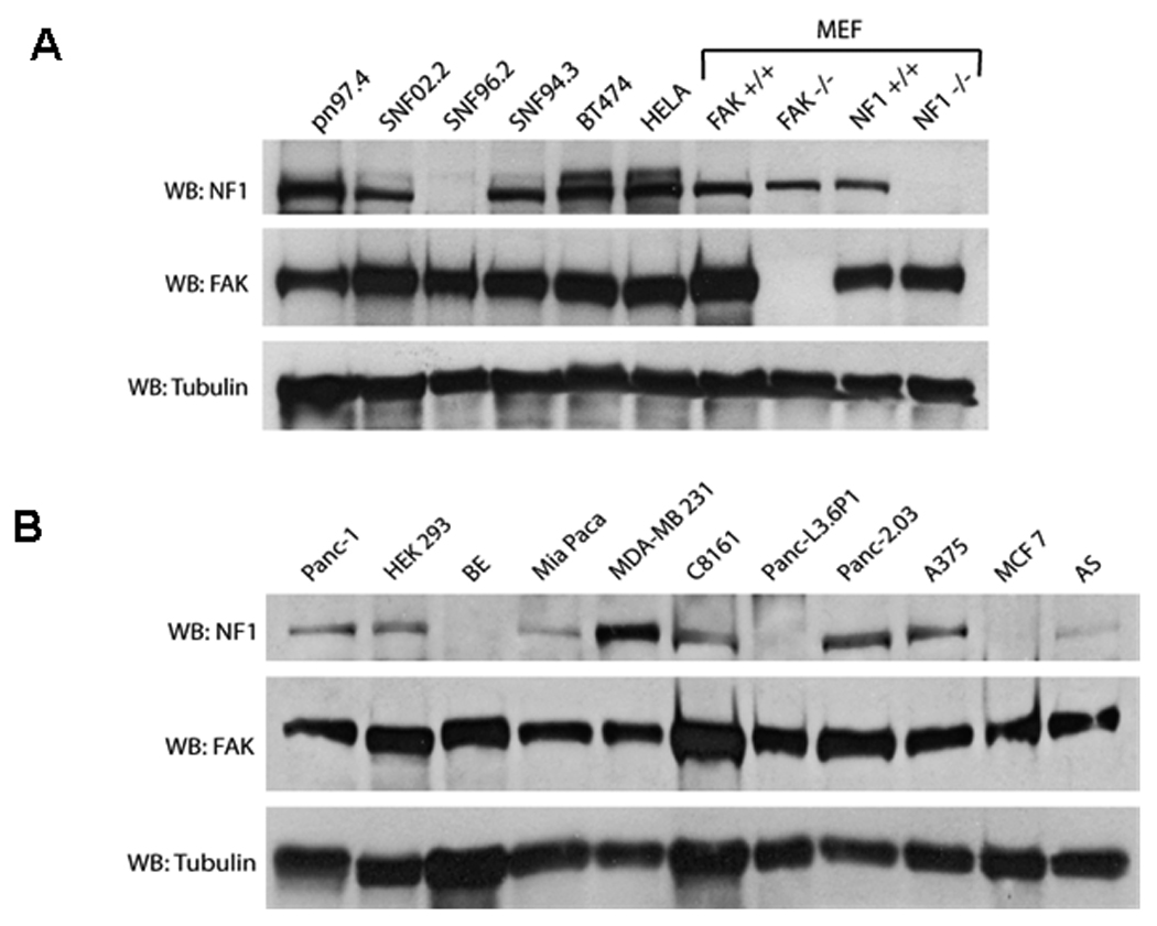
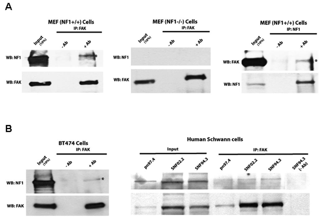
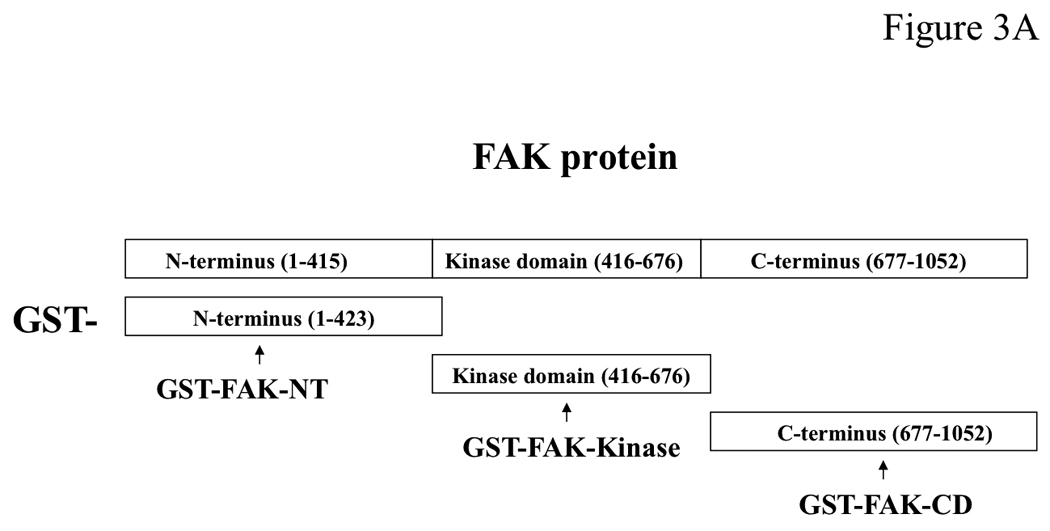
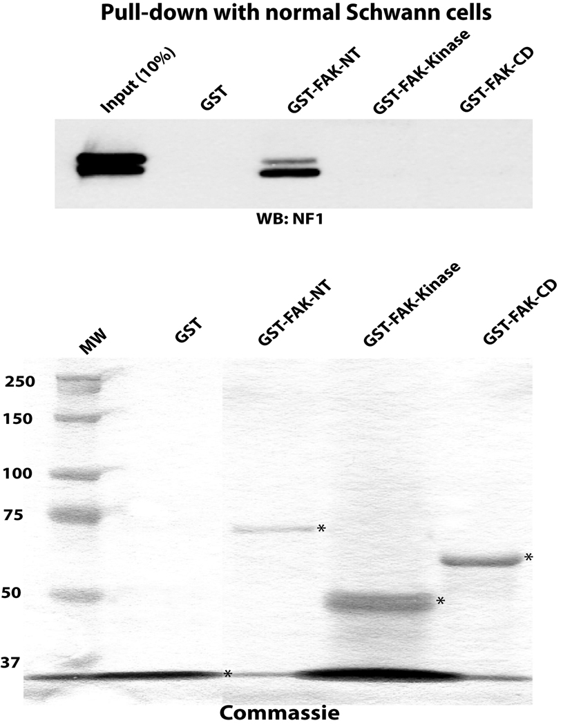
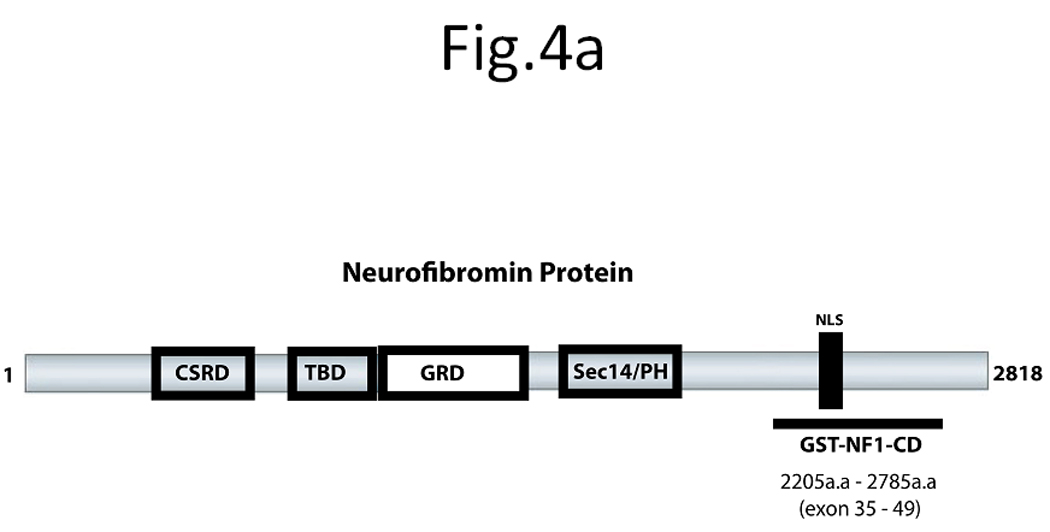
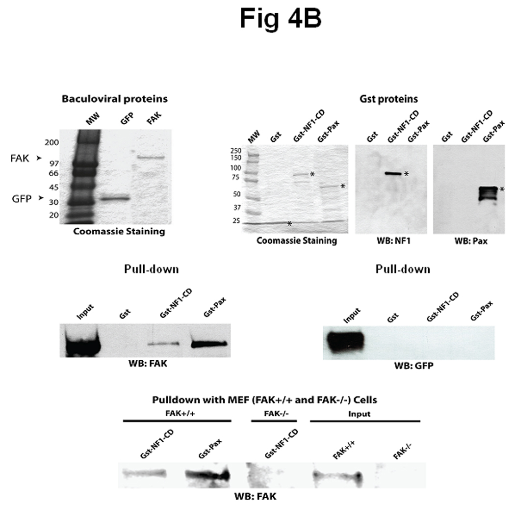
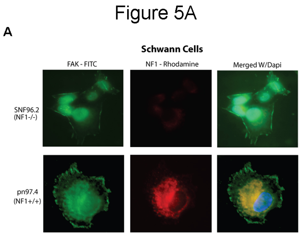
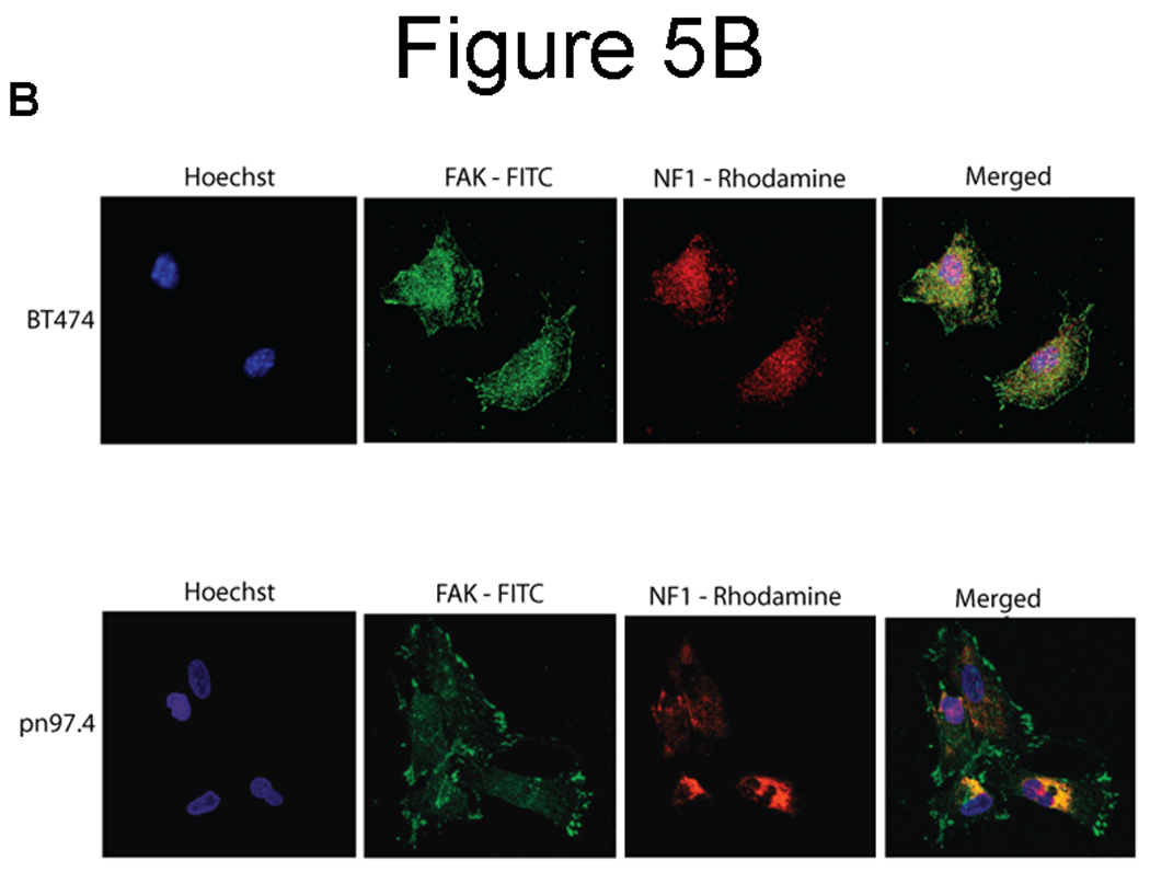
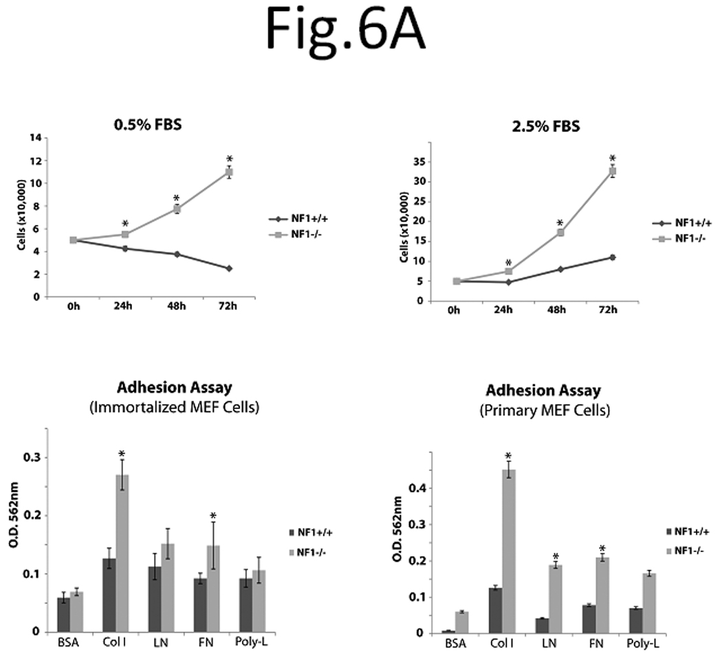
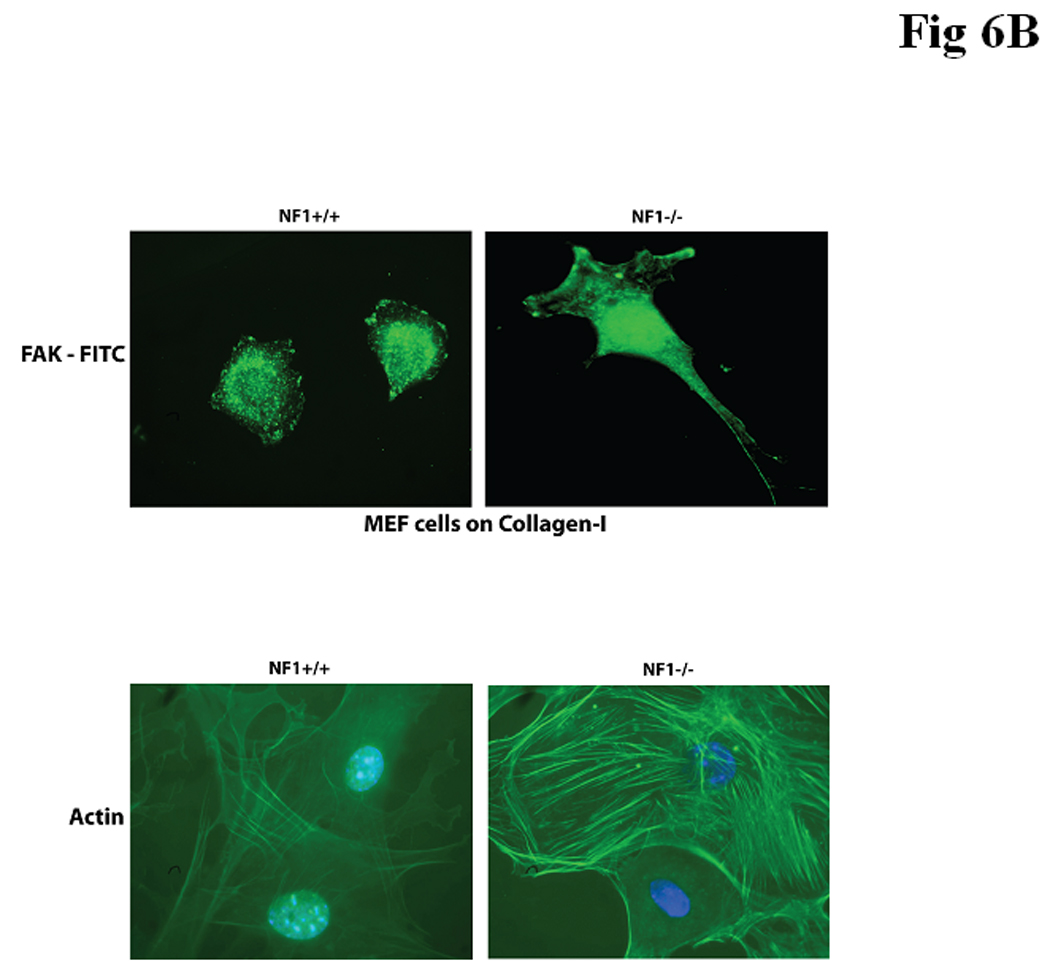
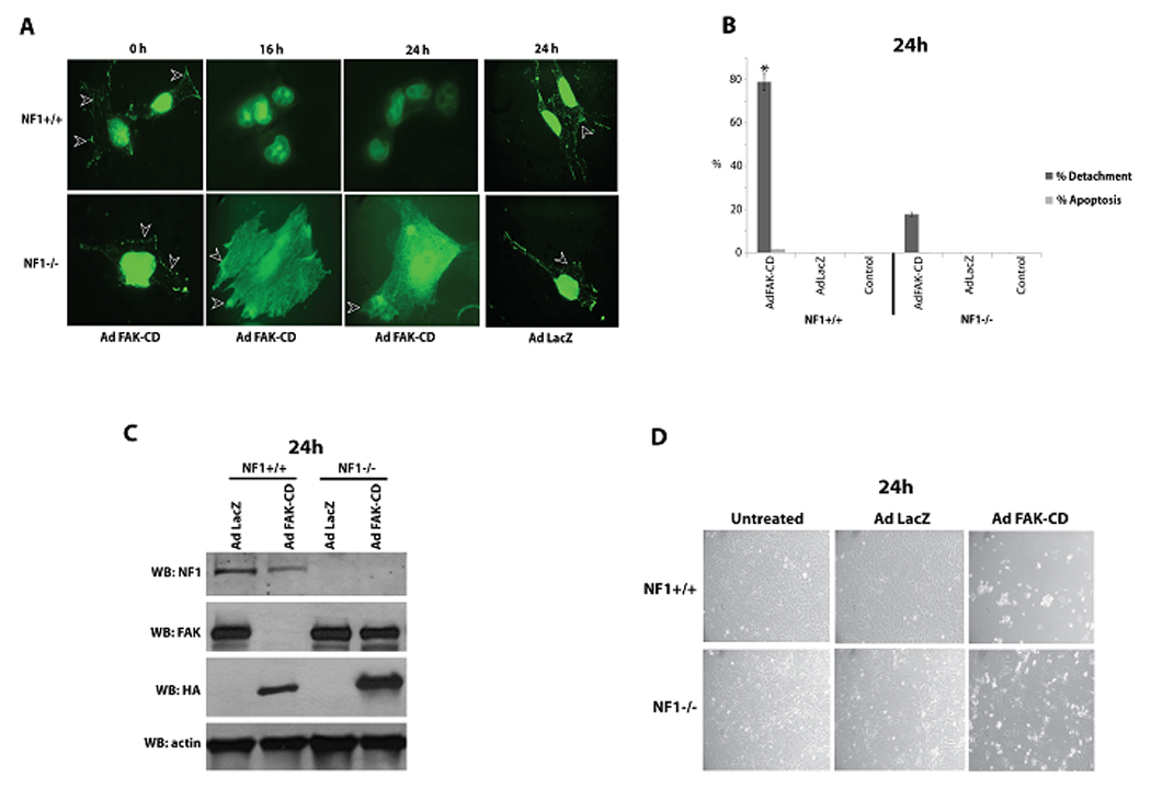
The NF1 gene that is altered in patients with type 1 neurofibromatosis (NF1) encodes a neurofibromin protein that functions as a tumor suppressor. In this report, we show for the first time physical interaction between neurofibromin and focal adhesion kinase (FAK), the protein that localizes at focal adhesions. We show that neurofibromin associates with the N-terminal domain of FAK, and that the C-terminal domain of neurofibromin directly interacts with FAK. Confocal microscopy demonstrates colocalization of NF1 and FAK in the cytoplasm, perinuclear and nuclear regions inside the cells. Nf1+/+ MEF cells expressed less cell growth during serum deprivation conditions, and adhered less on collagen and fibronectin-treated plates than Nf1(-/-) MEF cells, associated with changes in actin and FAK staining. In addition, Nf1+/+ MEF cells detached more significantly than Nf1(-/-) MEF cells by disruption of FAK signaling with the dominant-negative inhibitor of FAK, C-terminal domain of FAK (FAK-CD). Thus, the results demonstrate the novel interaction of neurofibromin and FAK and suggest their involvement in cell adhesion, cell growth, and other cellular events and pathways.


Figures

Figure 1
Figure 1. Expression of neurofibromin and FAK in different cancer cell lines
Western blotting analysis of neurofibromin and FAK expression was performed in normal Schwann cells (pn97.4), Malignant Peripheral Nerve Sheath Tumors (MPNST) (SNF02.2, SNF94.3, SNF96.2), breast cancer cells BT474, human cervical cancer HELA cells, mouse embryonic fibroblast (MEF) cells (FAK+/+ and FAK/, Nf1+/+ and Nf1/ (upper panel), and in pancreatic tumors (Panc-1, Mia Paca, Panc-L3.6p1, Panc-2.03), HEK 293 cells, neuroblastoma (BE, AS), melanoma (A375, C8161) and breast cancer MCF7, MDA-MB231 cells (lower panel).
Figure 2
Figure 2
Figure 2A. FAK and neurofibromin interact in Nf1+/+ cells in vivo. Left and middle panels: Immunoprecipitation of FAK was performed with anti-FAK C20 antibody followed by Western blotting with neurofibromin antibody in Nf1+/+ and Nf1/ MEF cells. Immunoprecipitation without antibody was used as a control (-Ab). Then membrane was stripped and Western blotting with FAK antibody was performed. FAK and NF1 interact in Nf1+/+, but not in Nf1/ MEF cells. Right panels: Immunoprecipitation with NF1 antibody detects interaction with FAK in Nf1+/+ MEF cells. Immunoprecipitation was performed with NF1 antibody, and then Western blotting with FAK antibody. Then membrane was stripped and Western blotting was performed with NF1 antibody. FAK and NF1 interacted in vivo in NF+/+ cells. Figure 2B. FAK and neurofibromin interact in breast cancer BT474 cells and in Human Schwann cells in vivo Immunoprecipitation with FAK antibody detected interaction with NF1 in BT474 (left panels) and in normal pn97.4 and Malignant Peripheral Nerve Sheath Tumors (MPNST) SNF02.2 and SNF94.3 Schwann cells (right panels). Immunoprecipitation was performed as above.
Figure 3
Figure 3. NF1 interacts with the N-terminal domain of FAK
A, The scheme of the GST FAK fusion proteins used for pull-down assay to demonstrate interaction of NF1 with FAK: GST-N-terminal FAK-NT; Central, FAK-Kinase and C-terminal FAK-CD domains. The GST proteins are marked by arrows. Upper panel: The three FAK domains (FAK-NT, FAK-Kinase and FAK-CD) were used in a pull-down assay with normal Schwann cells to identify which domain of FAK interacts with neurofibromin. After pull-down assay, Western blotting with the NF1 antibody was performed. The NF1 antibody detected of NF1 protein. Two bands of NF1 protein in normal Schwann cells were detected at high resolution gel electrophoresis conditions. Pull-down assay demonstrates interaction of NF1 with the N-terminal domain, but not with the Kinase or C-terminal domains of FAK. Lower panel: Coomassie staining of GST-FAK domain proteins. The GST-FAK protein domains were analyzed by Coomassie staining (lower panel, GST-proteins are marked by asterisks).
Figure 4
Figure 4. Direct interaction between FAK and neurofibromin in vitro
A) The scheme of neurofibromin domains structure. The scheme demonstrates the location of the recombinant C-terminal domain of NF1, GST-NF1-CD fusion protein (2205-2785 aa) inside neurofibromin protein, containing the CSRD (cysteine/serine Rich domain), TBD (tubulin binding domain, GRD (GAP-Related domain), Sec14 (sec14 domain) and NF1-NLS (nuclear localization signal). B) C-terminal domain of NF1 and FAK proteins directly interact in vitro. Left panel: Baculoviral purified FAK and GFP proteins were used for pull-down assay. Coomassie staining shows the fusion proteins that were confirmed by Western blotting with GFP and FAK antibodies [38]. Right panel: Coomassie staining and Western blotting analysis of GST, GST-NF1-CD and GST-paxillin fusion proteins (Materials and Methods) used in pull-down assay. Asterisks mark GST-fusion proteins. Western blotting with NF1 antibody and paxillin antibody confirmed NF1 and paxillin proteins. C). Pull-down with baculoviral FAK demonstrated direct binding with the C-terminal domain of NF1 protein. Left panel: Pull-down assay was performed with baculoviral FAK protein and GST, GST-NF1-CD and GST-paxillin. C-terminal domain of NF1 directly binds FAK protein. No binding of FAK with the negative control GST protein is detected, while it was detected with the positive control GST- paxilllin protein. Right panel: Pull-down with baculoviral GFP protein did not detect binding with FAK. The same pull-down as with baculoviral FAK protein was performed with baculoviral GFP protein. No binding of GST-NF1-CD was detected with GFP protein. D) Interaction of neurofibromin with FAK in FAK+/+, but not in FAK/ MEF cells. Pull-down assay of GST-NF1-CD protein and demonstrates interaction between GST-NF1-CD with endogenous FAK protein in FAK+/+, but not in FAK/ MEF cell extracts.
Figure 5
Figure 5. Co-localization of neurofibromin with FAK
A) Immunocytochemical and fluorescence microscopy analysis of FAK and neurofibromin localization in normal Schwann cells (pn97.4) (lower panel) and tumor Schwann cells (SNF96.2) lacking neurofibromin expression) (upper panel). FAK and neurofibromin co-localized in the cytoplasmic, peri-nuclear and nuclear regions in pn97.4 (Nf1+/+) cells. No co-localization is detected in SNF96.2 (Nf1/) cells. B) Confocal microscopy detects interaction of NF1 and FAK in breast cancer BT474 cells and pn97.4 Schwann cells. FAK and neurofibromin co-localization was observed in the cytoplasmic, peri-nuclear and nuclear regions in both cell lines.
Figure 6
Figure 6
Figure 6 A. Nf1+/+ MEF cells expressed significantly less cell growth at serum deprivation conditions and adhered less on Collagen I and Fibronectin compared with Nf1/ MEF cells. SV40 Immortalized Nf1+/+ and Nf1/ MEF cells were counted on hemocytometer at 0, 24, 48 and 72 hours after plating in medium with 0.5% FBS (upper left panel) or 2.5% FBS (upper right panel). Nf1+/+ MEF cells demonstrated significantly less cell growth at 0.5% and 2.5% serum versus Nf1/ MEF cells. Lower left panel: Adhesion of SV40 immortalized Nf1+/+ and Nf1/ MEF cells on BSA, Collagen I, Laminin, Fibronectin, and Poly-L lysine. SV40 immortalized Nf1+/+ and Nf1/ MEF cells were plated on BSA, Collagen I, Fibronectin, Laminin and Poly-L-lysine-coated plates, and adhesion assay was performed as described in Materials and Methods. Immortalized Nf1+/+ cells had significantly less adhesion on Collagen I and Fibronectin than Nf1/ cells. Lower right panel: Adhesion of normal primary Nf1+/+ and Nf1/ MEF cells on BSA, Collagen I, Laminin, Fibronectin, and Poly-L lysine The same experiment as with immortalized MEF cells (above) was performed with primary MEF fibroblasts. Primary Nf1+/+ cells had significantly less adhesion than Nf1/ cells. Figure 6B. FAK and actin staining in Nf1+/+ and Nf1/ MEF cells. Upper panel. FAK immunostaining was performed on immortalized Nf1+/+ and Nf1/ MEF cells, plated on Collagen I. Nf1/ and Nf1+/+ MEF cells had different cell morphology and FAK localization. Nf1/ cells had more elongated and more spread morphology and increased FAK staining at the leading edge of cells compared with less spread Nf1+/+ cells. Lower panel. Nf1/ MEF cells expressed increased actin staining compared with Nf1+/+ cells. Actin staining was performed with FITC-conjugated Phallacidin. Actin staining was increased in Nf1/ cells compared with Nf1+/+ MEF cells.
Figure 6
Figure 6
Figure 6 A. Nf1+/+ MEF cells expressed significantly less cell growth at serum deprivation conditions and adhered less on Collagen I and Fibronectin compared with Nf1/ MEF cells. SV40 Immortalized Nf1+/+ and Nf1/ MEF cells were counted on hemocytometer at 0, 24, 48 and 72 hours after plating in medium with 0.5% FBS (upper left panel) or 2.5% FBS (upper right panel). Nf1+/+ MEF cells demonstrated significantly less cell growth at 0.5% and 2.5% serum versus Nf1/ MEF cells. Lower left panel: Adhesion of SV40 immortalized Nf1+/+ and Nf1/ MEF cells on BSA, Collagen I, Laminin, Fibronectin, and Poly-L lysine. SV40 immortalized Nf1+/+ and Nf1/ MEF cells were plated on BSA, Collagen I, Fibronectin, Laminin and Poly-L-lysine-coated plates, and adhesion assay was performed as described in Materials and Methods. Immortalized Nf1+/+ cells had significantly less adhesion on Collagen I and Fibronectin than Nf1/ cells. Lower right panel: Adhesion of normal primary Nf1+/+ and Nf1/ MEF cells on BSA, Collagen I, Laminin, Fibronectin, and Poly-L lysine The same experiment as with immortalized MEF cells (above) was performed with primary MEF fibroblasts. Primary Nf1+/+ cells had significantly less adhesion than Nf1/ cells. Figure 6B. FAK and actin staining in Nf1+/+ and Nf1/ MEF cells. Upper panel. FAK immunostaining was performed on immortalized Nf1+/+ and Nf1/ MEF cells, plated on Collagen I. Nf1/ and Nf1+/+ MEF cells had different cell morphology and FAK localization. Nf1/ cells had more elongated and more spread morphology and increased FAK staining at the leading edge of cells compared with less spread Nf1+/+ cells. Lower panel. Nf1/ MEF cells expressed increased actin staining compared with Nf1+/+ cells. Actin staining was performed with FITC-conjugated Phallacidin. Actin staining was increased in Nf1/ cells compared with Nf1+/+ MEF cells.
Figure 7
Figure 7
A. Dominant-negative FAK inhibitor, Ad-FAK-CD causes displacement of FAK from focal adhesions in Nf1+/+ cells compared to Nf1−/− cells. Nf1+/+ and Nf1/ MEF cells were infected with Ad FAK-CD (300 FFU/cell) and Ad LacZ for 16 h and 24 h and cells were immunostained with anti-FAK 4.47 antibody to analyze focal adhesion formation (white arrowheads). Ad FAK-CD displaced FAK from focal adhesions in Nf1+/+ MEF cells but not in Nf1−/− MEF cells at 16h and at 24h. No displacement of FAK from focal adhesion was detected with control Ad-LacZ at 16 h (not shown) and at 24 h. Focal adhesion marked by white arrowheads. B. Detachment and apoptosis analysis of Nf1+/+ and Nf1/ MEF cells treated for 24 h with Ad FAK-CD. Detachment and apoptosis were detected, as described in Materials and Methods. Nf1+/+ cells had significantly more detachment in response to Ad-FAK-CD than Nf1/ MEF cells. C) FAK is displaced from focal adhesions by FAK-CD in Nf1+/+ cells compared with Nf1/ cells. Western blotting analysis of Ad-LacZ and Ad-FAK-CD-treated Nf1+/+ and Nf1/ MEF cells at 24 h. Western blotting with NF1, FAK, HA-tag and beta-Actin antibodies were performed. Western blotting with NF1 antibody shows NF1 expression in Nf1+/+ and absence in Nf1/ cells. Ad-FAK-CD caused degradation of FAK in Nf1+/+ cells, while Ad-LacZ did not cause this effect on FAK. NF−/ cells did not have FAK degradation in response to Ad-FAK-CD. Western blotting with HA-tag antibody demonstrates that both cell lines effectively express HAtagged FAK-CD. Western blotting with beta-actin show equal protein loading. D) Images under microscope of Nf1+/+ and Nf1/ MEF cells treated with Ad LacZ and Ad FAK-CD for 24 h. Ad-FAK-CD caused more detachment and cell rounding in Nf1+/+ cells compared with Nf1/ MEF cells.

Similar articles

Cited by

References

    1. Gutmann DH, Wood DL, Collins FS. Identification of the neurofibromatosis type 1 gene product. Proc Natl Acad Sci USA. 1991;88:9658–9662. - PMC - PubMed
    1. Wallace MR, Marchuk DA, Andersen LB, et al. Type 1 neurofibromatosis gene: identification of a large transcript disrupted in three NF1 patients. Science. 1990;249:181–189. - PubMed
    1. Marchuk DA, Saulino AM, Tavakkol R, et al. cDNA cloning of the type 1 neurofibromatosis gene: complete sequence of the NF1 gene product. Genomics. 1991;11:931–940. - PubMed
    1. Daston MM, Scrable H, Nordlund M, Sturbaum AK, Nissen LM, Ratner N. The protein product of the neurofibromatosis type 1 gene is expressed at highest abundance in neurons, Schwann cells, and oligodendrocytes. Neuron. 1992;8:415–428. - PubMed
    1. Scheffzek K, Ahmadian MR, Wiesmuller L, et al. Structural analysis of the GAP-related domain from neurofibromin and its implications. EMBO J. 1998;17:4313–4327. - PMC - PubMed

Publication types

Substances