. 2009 Nov 1;87(14):3076-87.
doi: 10.1002/jnr.22139.

Oxidative stress disrupts oligodendrocyte maturation

Affiliations

Oxidative stress disrupts oligodendrocyte maturation

Heather Morein French et al. J Neurosci Res. .

Abstract

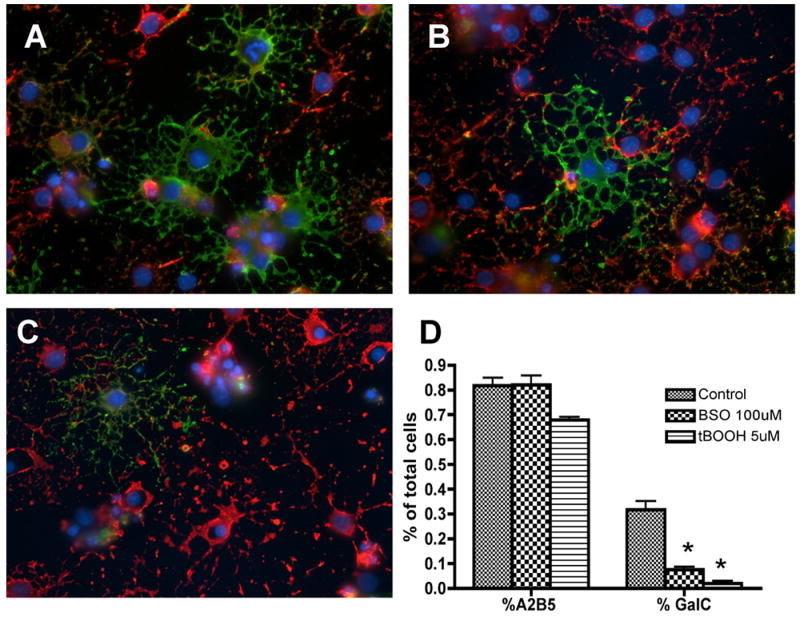
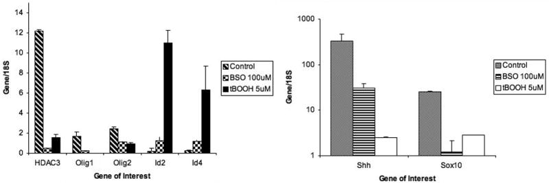
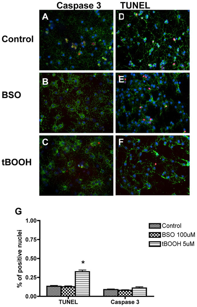
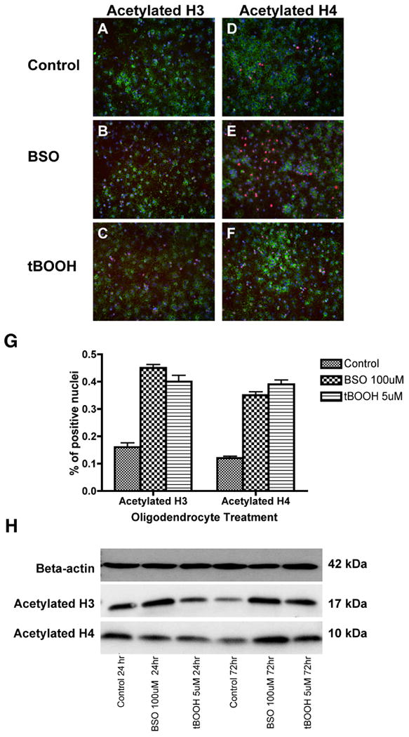
Periventricular white matter injury (PWMI) is the leading cause of chronic neurologic injury among survivors of preterm birth. The hallmark of PWMI is hypomyelination and a lack of mature, myelinating oligodendrocytes. Oligodendrocytes undergo a well-characterized lineage progression from neural stem cell to mature oligodendrocyte. Oligodendrocyte precursors have increased susceptibility to oxidative and free radical-mediated injury compared with mature oligodendrocytes as a result of lower levels of antioxidant enzymes and free radical scavengers. In this study, we show that oxidative stress disrupts oligodendrocyte differentiation by two mechanisms. First, oxidizing agents decrease the expression of key genes that promote oligodendrocyte differentiation from neural stem cells and increase the expression of genes known to inhibit differentiation. Second, global histone acetylation persists under conditions of oxidative stress, further contributing to the prevention of oligodendrocyte differentiation. Both of these mechanisms result in the arrest of oligodendrocyte differentiation without an increase in cell death.


Figures

Figure 1
Figure 1
Oligodendrocyte differentiation after 72 hours of oxidant exposure. OPCs were double-labeled in vitro with oligodendroglial markers A2B5 (red) and GalC (green) after 72 hours of oxidant exposure. The number of mature GalC+ cells was significantly decreased in the BSO (B) and tBOOH (C) exposed cultures compared to control (A) cultures. The majority of cells exposed to oxidative stress were arrested in the immature A2B5+ cell stage. Quantitative analysis of A2B5+ and GalC+ cells is shown in (D). The proportion of positive cells per 40× field was determined by counting 10 fields per coverslip from three separate experiments following indirect immunofluorescence using antibody to A2B5 and GalC. Data represent the mean plus standard deviation. *P < 0.05 vs. control.
Figure 2
Figure 2
Expression of genes promoting or inhibiting differentiation of oligodendrocyte precursor cells. The graph shows significantly decreased expression of Shh, Sox10, HDAC3, Olig1 and Olig2 72 hours after exposure to oxidants compared to control and significantly increased expression of Id2 and Id4 after exposure to oxidants. cDNA expression from 3 separate experiments, each performed in triplicate, was averaged. The data are presented as the mean ± standard deviation. *P < 0.05 vs. control.
Figure 3
Figure 3
Expression of Olig2 protein after exposure to oxidants is decreased. OPCs were labeled with Olig2 (red), A2B5 (green), and DAPI (blue) after 6 hours of oxidant exposure. Treatment with both tBOOH and BSO significantly reduced the number of cells strongly expressing Olig2. Arrows mark sample A2B5+ cells with decreased Olig2 expression.
Figure 4
Figure 4
Cell death was quantitated using both the TUNEL assay and activated caspase 3 staining following 72 hours of exposure to oxidative agents. The density of activated caspase 3+ (A-C) or TUNEL+ (D-F) nuclei was unchanged between control and BSO-exposed culture conditions. There is a small but significant increase in the number of TUNEL+ positive nuclei in the tBOOH-exposed cultures compared to control, but this increase doesn't appear to be large enough to account for the 10 to 20 fold decrease in key gene expression or decreased number of GalC+ oligodendrocytes. Quantitative analysis of activated caspase 3+ and TUNEL+ (G) cells is also shown. The proportion of positive cells per 40× field was determined by counting 10 fields per coverslip in 3 separate experiments following indirect immunofluorescence using antibody to activated caspase 3 or TUNEL analysis. Data represent the mean plus standard deviation. *P <0.05 vs. control.
Figure 5
Figure 5
OPCs were stained in vitro with antibodies to acetylated histone H3 (A-C) and acetylated histone H4 (D-F) after 72 hours of exposure to oxidative agents. The density of acetylated histone H3 and H4 was significantly higher in BSO- and tBOOH-exposed cells. Quantitative analysis of acetylated histone H3+ and H4+ nuclei is also shown (G). The proportion of positive cells per 20× field was determined by counting 10 fields per coverslip in three separate experiments following indirect immunofluorescence using antibody to acetylated histone H3 or H4. Data represent the mean plus standard deviation. *P <0.05. Western blotting was performed on OPC lysate after 24 and 72 hours (H). Analysis reveals an increase in acetylated histone H3 and H4 protein after 72 hours of exposure to oxidative stress. Each lane contains 25 μg protein.

Similar articles

Cited by

References

    1. Altman J, Bayer SA. The development of the rat spinal cord. In: Beck F, Hild W, van Limborgh J, Ortmann R, Pauly JE, Schiebler TH, editors. Advances in anatomy, embryology and cell biology. Vol. 85. Berlin: Springer Verlag; 1984. - PubMed
    1. Back SA, Gan XD, Li Y, Rosenberg PA, Volpe JJ. Maturation-dependent vulnerability of oligodendrocytes to oxidative stress-induced death caused by glutathione depletion. J Neurosci. 1998;18:6241–6253. - PMC - PubMed
    1. Back SA, Luo N, Borenstein N, Levine J, Volpe JJ, Kinney H. Late oligodendrocyte progenitors coincide with the developmental window of vulnerability for human perinatal white matter injury. J Neurosci. 2001;21:1302–1312. - PMC - PubMed
    1. Back SA, Volpe JJ, Kinney HH. Immunocytochemical characterization of oligodendrocyte development in human cerebral white matter. Soc Neurosci Abstr. 1996;20:1722.
    1. Ballas N, Battaglioli E, Atouf F, Andres ME, Chenoweth J, Anderson ME, Burger C, Moniwa M, Davie JR, Bowers WJ, Federoff HJ, Rose DW, Rosenfeld MG, Brehm P, Mandel G. Regulation of neuronal traits by a novel transcriptional complex. Neuron. 2001;31:353–365. - PubMed

Publication types