Review
. 2009 Nov;76(11):1019-32.
doi: 10.1002/mrd.21057.

Establishment of trophectoderm and inner cell mass lineages in the mouse embryo

Affiliations
Review

Establishment of trophectoderm and inner cell mass lineages in the mouse embryo

Yusuke Marikawa et al. Mol Reprod Dev. 2009 Nov.

Abstract

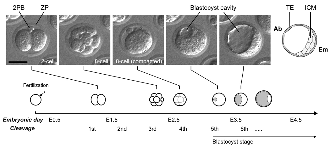
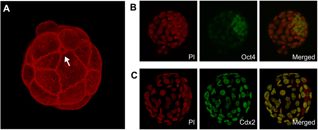
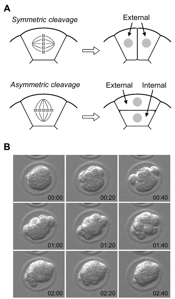
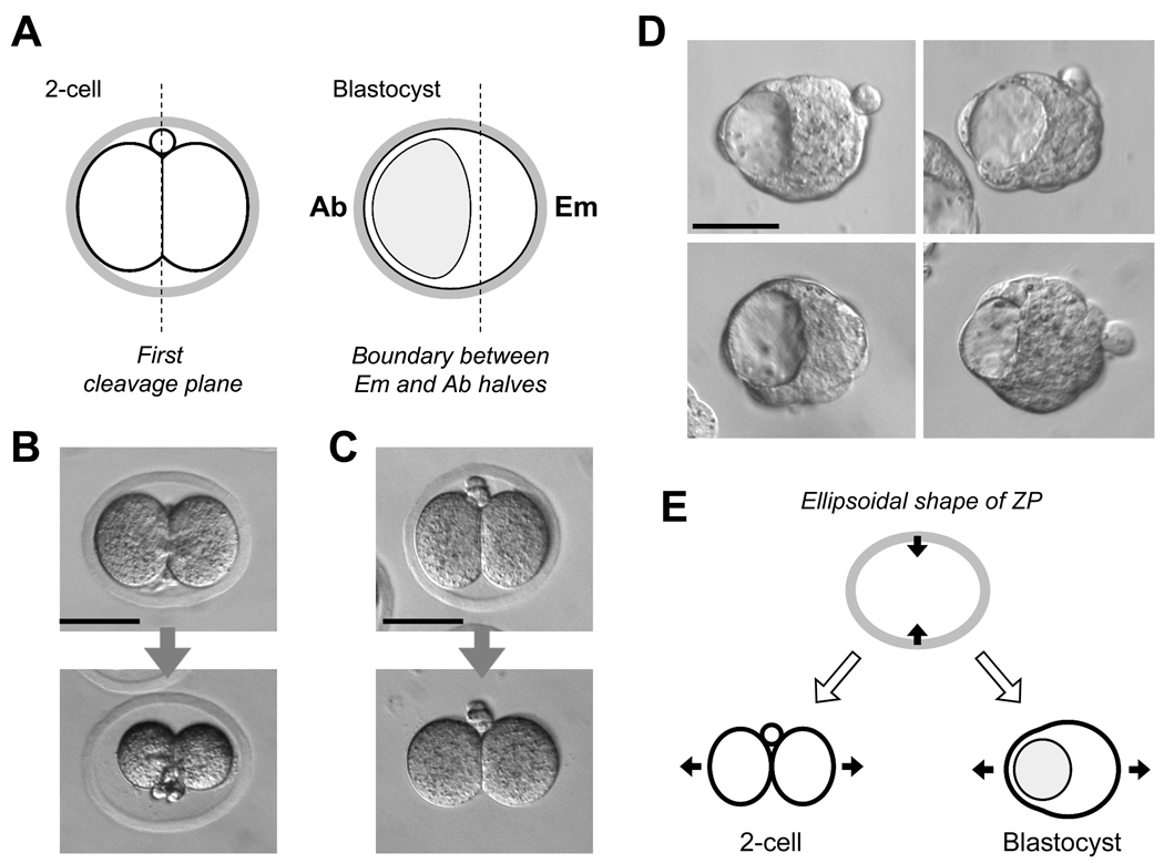
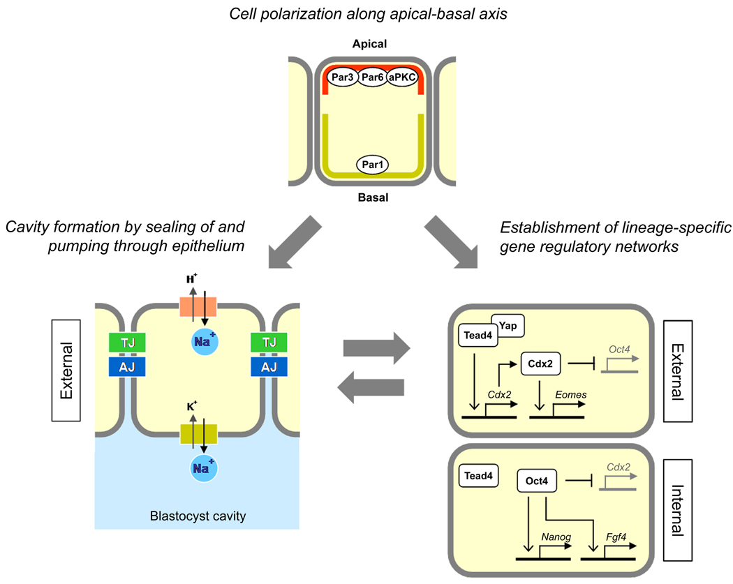
The first cell lineage specification in mouse embryo development is the formation of trophectoderm (TE) and inner cell mass (ICM) of the blastocyst. This article is to review and discuss the current knowledge on the cellular and molecular mechanisms of this particular event. Several transcription factors have been identified as the critical regulators of the formation or maintenance of the two cell lineages. The establishment of TE manifests as the formation of epithelium, and is dependent on many structural and regulatory components that are commonly found and that function in many epithelial tissues. Distinct epithelial features start to emerge at the late 8-cell stage, but the fates of blastomeres are not fixed as TE or ICM until around 32-cell stage. The location of blastomeres at this stage, that is, external or internal of the embryo, in effect defines the commitment towards the TE or ICM lineage, respectively. Some studies implicate the presence of a developmental bias among blastomeres at 2- or 4-cell stage, although it is unlikely to play a decisive role in the establishment of TE and ICM. The unique mode of cell lineage specification in the mouse embryo is further discussed in comparison with the formation of initial cell lineages, namely the three germ layers, in non-mammalian embryos.


Figures

Fig. 1
Fig. 1
Morphological transformation of mouse embryo during the first three days after fertilization. Embryonic day is often used in literatures to describe the developmental stage of embryos, which correspond to days after the mating of parents (or days post coitum). The timing of developmental progression in this diagram is based on embryos that are cultured in vitro. 2PB, second polar body; ZP, zona pellucida; TE, trophectoderm; ICM, inner cell mass; Ab, abembryonic side; Em, embryonic side. Scale bar, 50 µm.
Fig. 2
Fig. 2
(A) A 3D projection of confocal images of an embryo that is stained for actin filament using fluorescently labeled phalloidin. Belt-like intense staining of actin filament represents adherens junctions around the apical edge of cell-cell boundaries. The image is to highlight considerable variations among blastomeres in terms of the size of area that is exposed to the surface. Particularly, the one that is pointed out with an arrow has an extremely small surface exposed. This blastomere may be identified as either external or internal depending on methodologies used. (B) A 3D projection of confocal images of a late blastocyst that is stained for nuclei with propidium iodide (PI, red color) and for Oct4 protein with a specific antibody (green color). Note that Oct4 is most strongly expressed in the inner cell mass. (C) A 3D projection of confocal images of a late blastocyst that is stained for nuclei with PI and for Cdx2 protein with a specific antibody (green color). Note that Cdx2 is exclusively expressed in the trophectoderm.
Fig. 3
Fig. 3
(A) A schematic diagram depicting two types of cleavage patterns. Symmetric cleavage divides a blastomere along the apical-basal axis to generate two external blastomeres, whereas asymmetric cleavage divides perpendicular to the axis to generate one external and one internal blastomere. (B) Dynamic behaviors of blastomeres during cleavages can be seen by time-lapse cinematography. Images were taken every 20 minutes, starting at the beginning of the fourth cleavages (time is given in hours:minutes in each frame). Note that blastomeres during cleavages uncompact and then compact again, resulting in a dynamic change in the overall shape of the embryo.
Fig. 4
Fig. 4
(A) A schematic diagram depicting the relationship between the first cleavage plane at the 2-cell stage and the boundary between the embryonic and abembryonic halves at the blastocyst stage. (B) The ellipsoidal shape of zona pellucida (ZP) is stable and not dependent on the tension from the 2-cell stage blastomeres. When placed in a medium of high salt concentration, the embryo markedly shrunk, while the ZP maintained its elongated shape. (C) The ZP is firm and constricts an embryo within. The ZP was digested with pronase, which relieved an embryo from constriction. (D) Images of early blastocysts that were cultured without the ZP from the 8-cell stage. Note that embryos are slightly elongated along the Em-Ab axis likely due to the expansion of the blastocyst cavity. (E) A schematic diagram depicting how a mechanical constraint posed by an ellipsoidal ZP may affect the orientation of an elongated embryo at the 2-cell and blastocyst stages.
Fig. 5
Fig. 5
Summary diagrams depicting the roles of the molecules in the formation of TE and ICM that are discussed in this review article.

Similar articles

Cited by

References

    1. Alarcón VB, Marikawa Y. Deviation of the blastocyst axis from the first cleavage plane does not affect the quality of mouse postimplantation development. Biol Reprod. 2003;69:1208–1212. - PubMed
    1. Alarcón VB, Marikawa Y. Unbiased contribution of the first two blastomeres to mouse blastocyst development. Mol Reprod Dev. 2005;72:354–361. - PubMed
    1. Alarcón VB, Marikawa Y. Spatial alignment of the mouse blastocyst axis across the first cleavage plane is caused by mechanical constraint rather than developmental bias among blastomeres. Mol Reprod Dev. 2008;75:1143–1153. - PMC - PubMed
    1. Arnold SJ, Robertson EJ. Making a commitment: cell lineage allocation and axis patterning in the early mouse embryo. Nat Rev Mol Cell Biol. 2009;10:91–103. - PubMed
    1. Assémat E, Bazellières E, Pallesi-Pocachard E, Le Bivic A, Massey-Harroche D. Polarity complex proteins. Biochim Biophys Acta. 2008;1778:614–630. - PubMed

Publication types

Substances

LinkOut - more resources