

Neurons express hemoglobin alpha- and beta-chains in rat and human brains
- PMID: 19479992
- PMCID: PMC3123135
- DOI: 10.1002/cne.22062
Neurons express hemoglobin alpha- and beta-chains in rat and human brains
Abstract
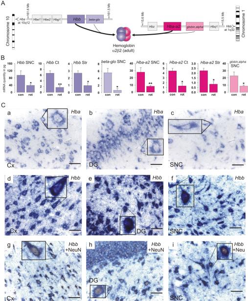
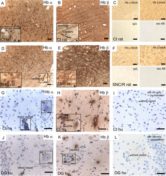
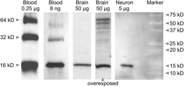
Hemoglobin is the oxygen carrier in vertebrate blood erythrocytes. Here we report that hemoglobin chains are expressed in mammalian brain neurons and are regulated by a mitochondrial toxin. Transcriptome analyses of laser-capture microdissected nigral dopaminergic neurons in rats and striatal neurons in mice revealed the presence of hemoglobin alpha, adult chain 2 (Hba-a2) and hemoglobin beta (Hbb) transcripts, whereas other erythroid markers were not detected. Quantitative reverse transcriptase-polymerase chain reaction (RT-PCR) analysis confirmed the expression of Hba-a2 and Hbb in nigral dopaminergic neurons, striatal gamma-aminobutyric acid (GABA)ergic neurons, and cortical pyramidal neurons in rats. Combined in situ hybridization histochemistry and immunohistochemistry with the neuronal marker neuronal nuclear antigen (NeuN) in rat brain further confirmed the presence of hemoglobin mRNAs in neurons. Immunohistochemistry identified hemoglobin alpha- and beta-chains in both rat and human brains, and hemoglobin proteins were detected by Western blotting in whole rat brain tissue as well as in cultures of mesencephalic neurons, further excluding the possibility of blood contamination. Systemic administration of the mitochondrial inhibitor rotenone (2 mg/kg/d, 7d, s.c.) induced a marked decrease in Hba-a2 and Hbb but not neuroglobin or cytoglobin mRNA in transcriptome analyses of nigral dopaminergic neurons. Quantitative RT-PCR confirmed the transcriptional downregulation of Hba-a2 and Hbb in nigral, striatal, and cortical neurons. Thus, hemoglobin chains are expressed in neurons and are regulated by treatments that affect mitochondria, opening up the possibility that they may play a novel role in neuronal function and response to injury.
Copyright 2009 Wiley-Liss, Inc.
Figures
References
-
- Agani FH, Pichiule P, Chavez JC, LaManna JC. The role of mitochondria in the regulation of hypoxia-inducible factor 1 expression during hypoxia. J Biol Chem. 2000;275:35863–35867. - PubMed
-
- Ajioka RS, Phillips JD, Kushner JP. Biosynthesis of heme in mammals. Biochim Biophys Acta. 2006;1763:723–736. - PubMed
-
- Bellelli A, Brunori M, Miele AE, Panetta G, Vallone B. The allosteric properties of hemoglobin: insights from natural and site directed mutants. Curr Protein Pept Sci. 2006;7:17–45. - PubMed
-
- Brazelton TR, Rossi FM, Keshet GI, Blau HM. From marrow to brain: expression of neuronal phenotypes in adult mice. Science. 2000;290:1775–1779. - PubMed
Publication types
MeSH terms
Substances
Grants and funding
LinkOut - more resources
Full Text Sources
Other Literature Sources
Miscellaneous
