. 2009 May 29:9:64.
doi: 10.1186/1471-2229-9-64.

In vivo reorganization of the actin cytoskeleton in leaves of Nicotiana tabacum L. transformed with plastin-GFP. Correlation with light-activated chloroplast responses

Affiliations

In vivo reorganization of the actin cytoskeleton in leaves of Nicotiana tabacum L. transformed with plastin-GFP. Correlation with light-activated chloroplast responses

Anna Anielska-Mazur et al. BMC Plant Biol. .

Abstract

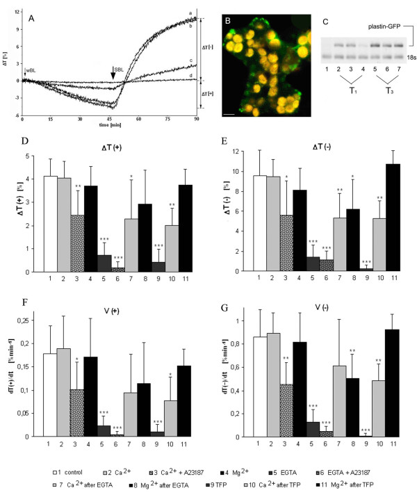
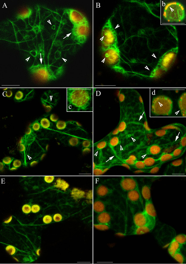
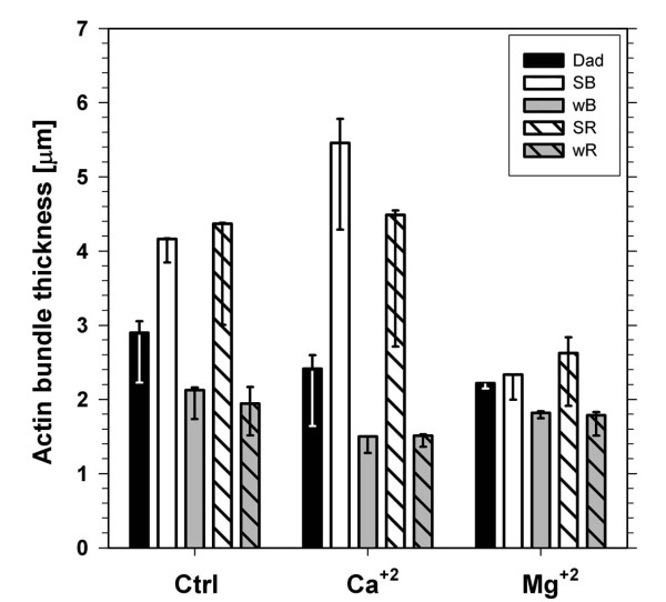
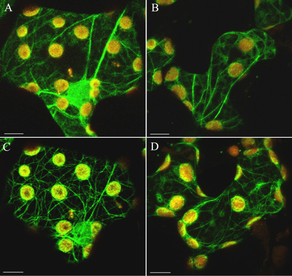
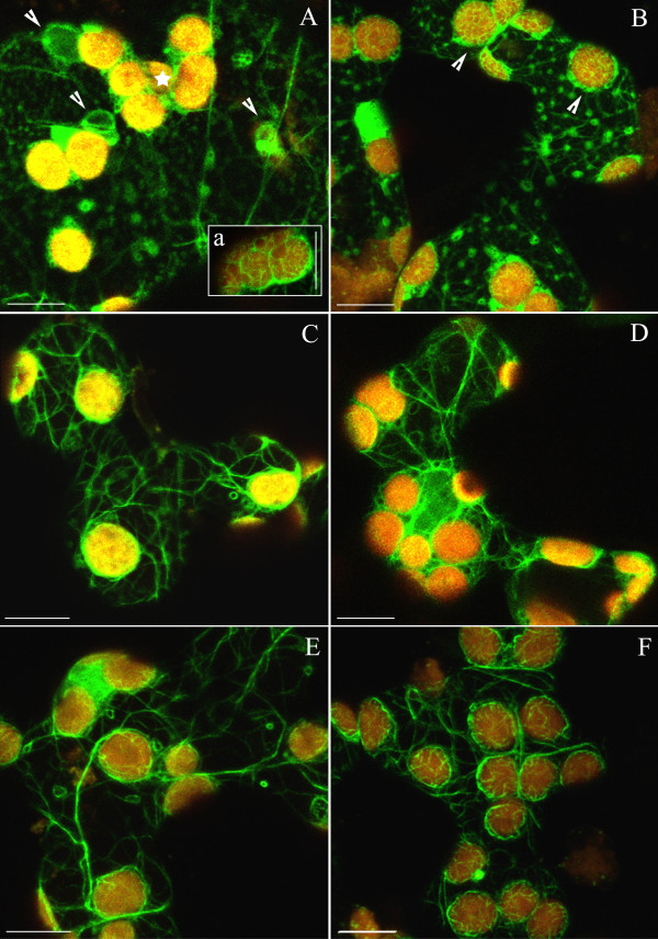
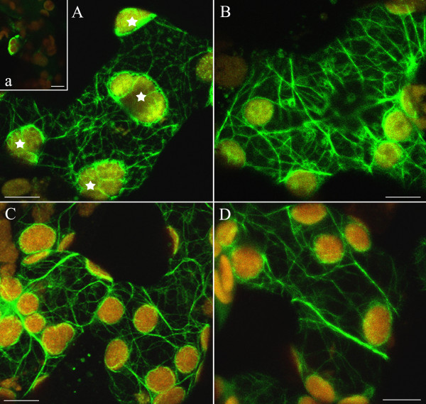
Background: The actin cytoskeleton is involved in the responses of plants to environmental signals. Actin bundles play the role of tracks in chloroplast movements activated by light. Chloroplasts redistribute in response to blue light in the mesophyll cells of Nicotiana tabacum. The aim of this work was to study the relationship between chloroplast responses and the organization of actin cytoskeleton in living tobacco cells. Chloroplast movements were measured photometrically as changes in light transmission through the leaves. The actin cytoskeleton, labeled with plastin-GFP, was visualised by confocal microscopy.

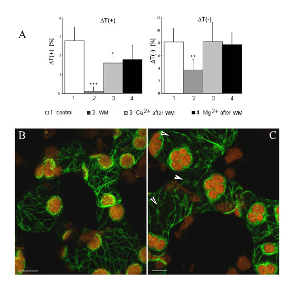
Results: The actin cytoskeleton was affected by strong blue and red light. No blue light specific actin reorganization was detected. EGTA and trifluoperazine strongly inhibited chloroplast responses and disrupted the integrity of the cytoskeleton. This disruption was reversible by Ca(2+) or Mg(2+). Additionally, the effect of trifluoperazine was reversible by light. Wortmannin, an inhibitor of phosphoinositide kinases, potently inhibited chloroplast responses but did not influence the actin cytoskeleton at the same concentration. Also this inhibition was reversed by Ca(2+) and Mg(2+). Magnesium ions were equally or more effective than Ca(2+) in restoring chloroplast motility after treatment with EGTA, trifluoperazine or wortmannin.

Conclusion: The architecture of the actin cytoskeleton in the mesophyll of tobacco is significantly modulated by strong light. This modulation does not affect the direction of chloroplast redistribution in the cell. Calcium ions have multiple functions in the mechanism of the movements. Our results suggest also that Mg(2+) is a regulatory molecule cooperating with Ca(2+) in the signaling pathway of blue light-induced tobacco chloroplast movements.


Figures

Figure 1
Figure 1
(A) Chloroplast responses to blue light in wild-type and transgenic N. tabacum cv. Samsun expressing plastin-GFP. The curves show changes in transmission of red measuring light (ΔT) through dark-adapted leaves exposed to continuous weak blue light (wBL, 0.4 Wm-2, 45 min) and strong blue light (SBL, 10 Wm-2, 45 min). ΔT [+] and ΔT [-] denote amplitudes of accumulation and avoidance responses respectively. Representative responses for about seventy tests carried out with three-month-old wild-type (a) and transgenic (b) tobacco plants. Curves c and d are representative of two-month (c) and one-month-old (d) plants. (B) Actin organization in immature mesophyll of transgenic (one-month-old) plants grown in vitro. Bar, 10 μm. (C) RT-PCR. (1) control, non-transformed plant, (2–4) three plants of T1 generation, (5–7) three plants of T3 generation. (D – G) Parameters of blue light-controlled chloroplast responses in mature leaves of transgenic N. tabacum. (D, E) Amplitudes: ΔT(+) of weak (wBL, 0.4 Wm-2), and ΔT(-) of strong (SBL, 10 Wm-2) blue light responses. (F, G) Velocities: V(+) of wBL, and V(-) of SBL responses. Averages of 7–14 measurements. Error bars represent SD. Asterisks denote the significance of differences (p-value calculated with the unpaired t-test, * p = 0,05–0,001; ** p = 0,001–0,0001; *** p < 0,0001).
Figure 2
Figure 2
The actin cytoskeleton in 3-month-old tobacco mesophyll cells as visualized by plastin-GFP (green fluorescence). (A, B) Network of actin in dark-adapted cells. "Baskets" around chloroplasts marked with arrows and circular structures marked with arrowheads; yellow-red colour comes from autofluorescence of chloroplasts. (C) Reorganization of F-actin after 1 h of exposure to continuous wBL (0.4 Wm-2). Strands which spread across the cortical cytoplasm, frequently split into thinner filaments (marked with arrowheads). (c) Actin filaments forming baskets are better resolved after wBL. (D) Wide bands of F-actin (arrows) have a loose contact with chloroplasts after exposure to SBL (10 Wm-2, 20 min). (E) Effect of continuous wRL (0.24 Wm-2, 1 h) or (F) to SRL (6.7 Wm-2, 20 min) on the actin cytoskeleton. Scale bars, 10 μm. The cytoskeleton forms numerous small loops (B and D, arrowheads), most of them containing mitochondria. Insets b and d show magnified chloroplasts with mitochondria visible (orange/red colour) in the AC loops after staining with TMRE.
Figure 3
Figure 3
Thickness of actin bundles in control (Ctrl) and in the presence of calcium (Ca+2) and magnesium (Mg+2) ions. The bundle thickness was measured in cells adapted to darkness (Dad, black bars) and in cells illuminated with strong (SB) or weak (wB) blue light (empty and gray bars, respectively) and with strong (SR) or weak (wR) red light (backslashed and backslashed gray bars, respectively). The thickness was calculated in micrometers (μm) as 99th percentile of the data corresponding to averages of optical sections, whereas error bars represent 95% confidence intervals.
Figure 4
Figure 4
Influence of calcium and magnesium ions on the actin network in dark-adapted and weak light-irradiated cells. Samples were incubated for 2 h with 5 mM Ca2+ (A, C) or 5 mM Mg2+ (B, D). Actin cytoskeleton in the dark-adapted cells (A, B) and after irradiation with continuous weak red light for 1 h (C, D). Scale bars, 10 μm.
Figure 5
Figure 5
Disintegration of F-actin in EGTA and restoration of actin network prompted by calcium or magnesium ions. (A) Formation of actin foci in response to 0,5–1 h incubation with 1 mM EGTA in dark-adapted cells. Fluorescent spots and loops of various sizes (arrowheads) are visible throughout the cytoplasm. Chloroplasts are arranged into tight clusters (asterisk). Thin filaments are present on chloroplast surfaces (a). (B) Actin foci persist after wBL irradiation. Distinct baskets around chloroplasts (arrowheads) became more visible after exposure to weak light. Effect of 5 mM Ca2+ (C, D) or 5 mM Mg2+ (E, F), each applied for 2 h on actin organization in EGTA pre-treated cells. In both cases, F-actin network recovered in dark-adapted cells (C, E) and after additional exposure to continuous wBL for 1 h (D, F). Scale bars, 10 μm.
Figure 6
Figure 6
Disintegration of actin bundles by trifluoperazine and its reversal by Ca2+ and Mg2+.(A) Images of disordered F-actin after treatment with 20 μM TFP for 30 min; chloroplast clusters marked with asterisks. Inset (a): effect of 1 h treatment with TFP in darkness. (B) Recovery of actin bundles by continuous wBL (1 h). The irradiation started 15 min after the onset of TFP treatment. The complete AC reconstruction in dark-adapted mesophyll cells pre-treated with TFP for 30 min and thereafter incubated with 5 mM Ca2+ or 5 mM Mg2+ for 2 h (C, D, respectively). Scale bars, 10 μm.
Figure 7
Figure 7
Effect of wortmannin (WM) on BL-induced chloroplast responses and on the actin cytoskeleton in tobacco leaves. (A) Amplitudes: ΔT(+) of weak (wBL, 0.4 Wm-2), and ΔT(-) of strong (SBL, 10 Wm-2) blue light-induced responses after 1.5 h incubation with 10 μM WM. The leaves were subsequently treated with 5 mM Ca2+ or Mg2+ for 3 h (right columns). Averages of 5–7 measurements. Error bars represent SD. Asterisks denote the significance of differences (p-value calculated with the unpaired t-test, * p = 0,05–0,001; ** p = 0,001–0,0001; *** p < 0,0001). The network of actin bundles in the mesophyll cells of the transgenic tobacco after 1,5 h incubation with 10 μM (B), and 50 μM (C) WM. Note the chloroplast clustering and disturbances of AC integrity (arrowheads) at the higher WM concentration. Scale bars, 10 μm.

Similar articles

Cited by

References

    1. Grolig F. Organelle movements: transport and positioning. In: Hussey PJ, editor. The plant cytoskeleton in cell differentiation and development. Oxford, Blackwell Publishing; 2004. pp. 148–175.
    1. Drøbak BK, Franklin-Tong VE, Staiger CJ. The role of the actin cytoskeleton in plant cell signaling. New Phytologist. 2004;163:13–30. doi: 10.1111/j.1469-8137.2004.01076.x. - DOI - PubMed
    1. Haupt W, Scheuerlein R. Chloroplast movement. Plant Cell Environ. 1990;13:596–614. doi: 10.1111/j.1365-3040.1990.tb01078.x. - DOI
    1. Sakamoto K, Briggs WR. Cellular and subcellular localization of phototropin 1. Plant Cell. 2002;14:1723–1735. doi: 10.1105/tpc.003293. - DOI - PMC - PubMed
    1. Harada A, Sakai T, Okada K. Phot1 and phot2 mediate blue light-induced transient increase in cytosolic Ca2+ differently in Arabidopsis leaves. Proc Natl Acad Sci USA. 2003;100:8583–8588. doi: 10.1073/pnas.1336802100. - DOI - PMC - PubMed

Publication types

MeSH terms

LinkOut - more resources