. 2009 Sep;30(25):4094-103.
doi: 10.1016/j.biomaterials.2009.04.024. Epub 2009 May 23.

Phase separation, pore structure, and properties of nanofibrous gelatin scaffolds

Affiliations

Phase separation, pore structure, and properties of nanofibrous gelatin scaffolds

Xiaohua Liu et al. Biomaterials. 2009 Sep.

Abstract

The development of three-dimensional (3D) biomimetic scaffolds which provide an optimal environment for cells adhesion, proliferation and differentiation, and guide new tissue formation has been one of the major goals in tissue engineering. In this work, a processing technique has been developed to create 3D nanofibrous gelatin (NF-gelatin) scaffolds, which mimic both the physical architecture and the chemical composition of natural collagen. Gelatin matrices with nanofibrous architecture were first created by using a thermally induced phase separation (TIPS) technique. Macroporous NF-gelatin scaffolds were fabricated by combining the TIPS technique with a porogen-leaching process. The processing parameters were systematically investigated in relation to the fiber diameter, fiber length, surface area, porosity, pore size, interpore connectivity, pore wall architecture, and mechanical properties of the NF-gelatin scaffolds. The resulting NF-gelatin scaffolds possess high surface areas (>32 m(2)/g), high porosities (>96%), well-connected macropores, and nanofibrous pore wall structures. The technique advantageously controls macropore shape and size by paraffin spheres, interpore connectivity by assembly conditions (time and temperature of heat treatment), pore wall morphology by phase separation and post-treatment parameters, and mechanical properties by polymer concentration and crosslinking density. Compared to commercial gelatin foam (Gelfoam), the NF-gelatin scaffold showed much better dimensional stability in a tissue culture environment. The NF-gelatin scaffolds, therefore, are excellent scaffolds for tissue engineering.


Figures

Figure 1
Figure 1
SEM micrographs of gelatin matrices fabricated from different solvent mixtures and processing conditions: (a) water, lyophilized; (b) high magnification of (a); (c) ethanol/water = 50/50, phase separated at −76°C, solvent exchanged, and freeze-dried; (d) methanol/water = 30/70, phase separated at −76°C, solvent exchanged, and freeze-dried. The gelatin concentration was 5.0% (wt/v) for all samples.
Figure 1
Figure 1
SEM micrographs of gelatin matrices fabricated from different solvent mixtures and processing conditions: (a) water, lyophilized; (b) high magnification of (a); (c) ethanol/water = 50/50, phase separated at −76°C, solvent exchanged, and freeze-dried; (d) methanol/water = 30/70, phase separated at −76°C, solvent exchanged, and freeze-dried. The gelatin concentration was 5.0% (wt/v) for all samples.
Figure 2
Figure 2
SEM micrographs of gelatin matrices fabricated using ethanol/water solvent mixtures. (a) ethanol/water = 10/90 (v/v); (b) ethanol/water = 20/80 (v/v); (c) ethanol/water = 60/40 (v/v). The gelatin concentration was 5.0% (wt/v) for all samples.
Figure 2
Figure 2
SEM micrographs of gelatin matrices fabricated using ethanol/water solvent mixtures. (a) ethanol/water = 10/90 (v/v); (b) ethanol/water = 20/80 (v/v); (c) ethanol/water = 60/40 (v/v). The gelatin concentration was 5.0% (wt/v) for all samples.
Figure 2
Figure 2
SEM micrographs of gelatin matrices fabricated using ethanol/water solvent mixtures. (a) ethanol/water = 10/90 (v/v); (b) ethanol/water = 20/80 (v/v); (c) ethanol/water = 60/40 (v/v). The gelatin concentration was 5.0% (wt/v) for all samples.
Figure 3
Figure 3
SEM micrographs of SW-gelatin scaffolds and NF-gelatin scaffolds. (a) SW-gelatin scaffold, ×50; (b) pore wall morphology of SW-gelatin scaffold, ×1000; (c) NF-gelatin scaffold, ×50; (d) pore wall morphology of NF-gelatin scaffold, ×1000; (e) high magnification of (d), ×10000. Scaffolds were prepared from a 7.5% (wt/v) gelatin solution in ethanol/water mixture (ethanol/water = 50/50) and the paraffin spheres were 250–420 μm in diameter. Paraffin spheres were heat treated at 37°C for 40 min.
Figure 3
Figure 3
SEM micrographs of SW-gelatin scaffolds and NF-gelatin scaffolds. (a) SW-gelatin scaffold, ×50; (b) pore wall morphology of SW-gelatin scaffold, ×1000; (c) NF-gelatin scaffold, ×50; (d) pore wall morphology of NF-gelatin scaffold, ×1000; (e) high magnification of (d), ×10000. Scaffolds were prepared from a 7.5% (wt/v) gelatin solution in ethanol/water mixture (ethanol/water = 50/50) and the paraffin spheres were 250–420 μm in diameter. Paraffin spheres were heat treated at 37°C for 40 min.
Figure 4
Figure 4
SEM micrographs of 3D NF-gelatin scaffolds with varying macropore sizes: (a) 150–250 μm; (b) 250–420 μm; (c) 420–600 μm. Scaffolds were prepared from a 7.5% (wt/v) gelatin solution in ethanol/water mixture (ethanol/water = 50/50) and the paraffin spheres were heat treated at 37°C for 30 min.
Figure 4
Figure 4
SEM micrographs of 3D NF-gelatin scaffolds with varying macropore sizes: (a) 150–250 μm; (b) 250–420 μm; (c) 420–600 μm. Scaffolds were prepared from a 7.5% (wt/v) gelatin solution in ethanol/water mixture (ethanol/water = 50/50) and the paraffin spheres were heat treated at 37°C for 30 min.
Figure 5
Figure 5
SEM micrographs of 3D NF-gelatin scaffolds with varying interpore connectivities under different heat treatment conditions: (a) 37°C for 20 min; (b) 37°C for 50 min; (c) 37°C for 200 min; (d) 37°C for 400 min. Scaffolds were prepared from a 7.5% (wt/v) gelatin solution in ethanol/water mixture (ethanol/water = 50/50) and the paraffin spheres were 250–420 μm in diameter.
Figure 5
Figure 5
SEM micrographs of 3D NF-gelatin scaffolds with varying interpore connectivities under different heat treatment conditions: (a) 37°C for 20 min; (b) 37°C for 50 min; (c) 37°C for 200 min; (d) 37°C for 400 min. Scaffolds were prepared from a 7.5% (wt/v) gelatin solution in ethanol/water mixture (ethanol/water = 50/50) and the paraffin spheres were 250–420 μm in diameter.
Figure 6
Figure 6
SEM micrographs of pore wall morphology of gelatin scaffolds before and after crosslinking in different solvent systems. (a) before crosslinking; (b) after crosslinking in aqueous EDC solution; (c) after crosslinking in EDC/Acetone/water solution (acetone/water = 90/10, v/v). Scaffolds were prepared from a 10.0% (wt/v) gelatin solution in ethanol/water mixture (ethanol/water = 50/50) and the paraffin spheres were 250–420 μm in diameter.
Figure 6
Figure 6
SEM micrographs of pore wall morphology of gelatin scaffolds before and after crosslinking in different solvent systems. (a) before crosslinking; (b) after crosslinking in aqueous EDC solution; (c) after crosslinking in EDC/Acetone/water solution (acetone/water = 90/10, v/v). Scaffolds were prepared from a 10.0% (wt/v) gelatin solution in ethanol/water mixture (ethanol/water = 50/50) and the paraffin spheres were 250–420 μm in diameter.
Figure 7
Figure 7
Swelling volume ratios in water after chemical crosslinking in acetone/water and dioxane/water systems. Scaffolds were prepared using 7.5% (wt/v) gelatin solution; the pore size was 250–420 μm. EDC concentration was 5 mmol/L, EDC/gelatin = 5/1 (mg/mg).
Figure 8
Figure 8
(a) The compressive modulus comparison of Gelfoam®, NF-gelatin and SW-gelatin scaffolds. The NF-gelatin and SW-gelatin scaffolds were prepared from a 7.5% (wt/v) gelatin solution in an ethanol/water mixture (ethanol/water = 50/50), had a macropore size of 250–420 μm and a porosity of about 97.5%. (b) The effects of gelatin concentration on the compressive modulus of NF-gelatin scaffolds. The NF-gelatin scaffolds had a macropore size ranging of 250–420 μm. (c) The effect of macropore size on compressive modulus of both NF-gelatin scaffolds and SW-gelatin scaffolds. The NF-gelatin and SW-gelatin scaffolds were prepared from a 7.5% (wt/v) gelatin solution in an ethanol/water mixture (ethanol/water = 50/50).
Figure 8
Figure 8
(a) The compressive modulus comparison of Gelfoam®, NF-gelatin and SW-gelatin scaffolds. The NF-gelatin and SW-gelatin scaffolds were prepared from a 7.5% (wt/v) gelatin solution in an ethanol/water mixture (ethanol/water = 50/50), had a macropore size of 250–420 μm and a porosity of about 97.5%. (b) The effects of gelatin concentration on the compressive modulus of NF-gelatin scaffolds. The NF-gelatin scaffolds had a macropore size ranging of 250–420 μm. (c) The effect of macropore size on compressive modulus of both NF-gelatin scaffolds and SW-gelatin scaffolds. The NF-gelatin and SW-gelatin scaffolds were prepared from a 7.5% (wt/v) gelatin solution in an ethanol/water mixture (ethanol/water = 50/50).
Figure 9
Figure 9
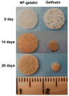
The size variation of scaffolds (NF-gelatin and Gelfoam®) after culturing MC3T3-E1 osteoblasts for 2 weeks and 4 weeks. NF-gelatin scaffolds were prepared from a 7.5% (wt/v) gelatin solution in an ethanol/water mixture (ethanol/water = 50/50) with a macropore size of 250–420 μm.

References

    1. Langer R, Vacanti JP. Tissue Engineering. Science. 1993;260(5110):920–926. - PubMed
    1. Ma PX. Biomimetic materials for tissue engineering. Advanced Drug Delivery Reviews. 2008;60(2):184–198. - PMC - PubMed
    1. Kadler KE, Holmes DF, Trotter JA, Chapman JA. Collagen fibril formation. Biochemical Journal. 1996;316:1–11. - PMC - PubMed
    1. Elsdale T, Bard J. Collagen Substrata for Studies on Cell Behavior. Journal of Cell Biology. 1972;54(3):626. - PMC - PubMed
    1. Eyre DR. Collagen - Molecular Diversity in the Bodys Protein Scaffold. Science. 1980;207(4437):1315–1322. - PubMed

Publication types