. 2009 Jul;33(7):1109-32.
doi: 10.1016/j.neubiorev.2009.05.005. Epub 2009 May 27.

The dopaminergic basis of human behaviors: A review of molecular imaging studies

Affiliations

The dopaminergic basis of human behaviors: A review of molecular imaging studies

Alice Egerton et al. Neurosci Biobehav Rev. 2009 Jul.

Abstract

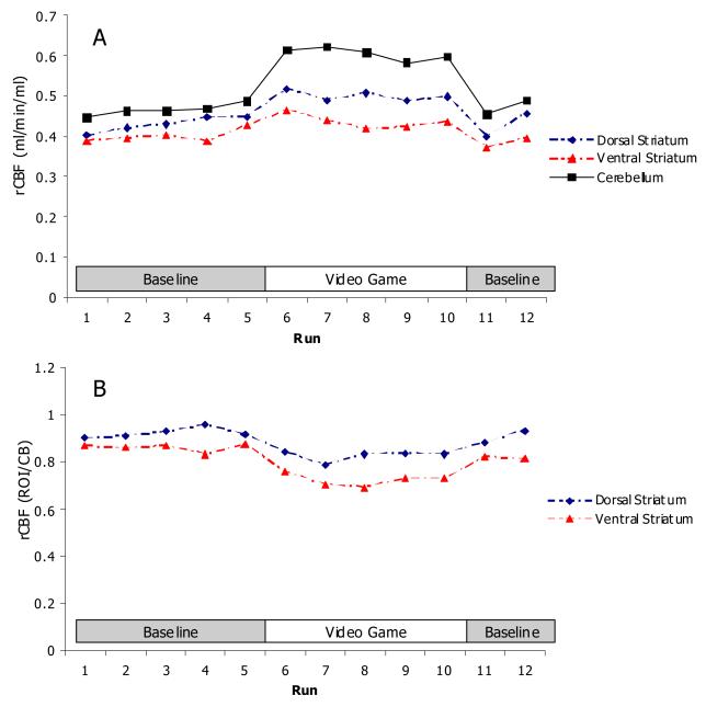
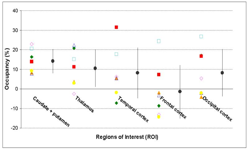
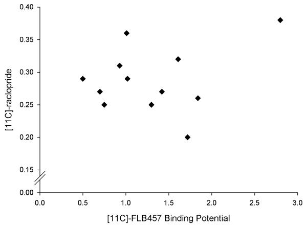
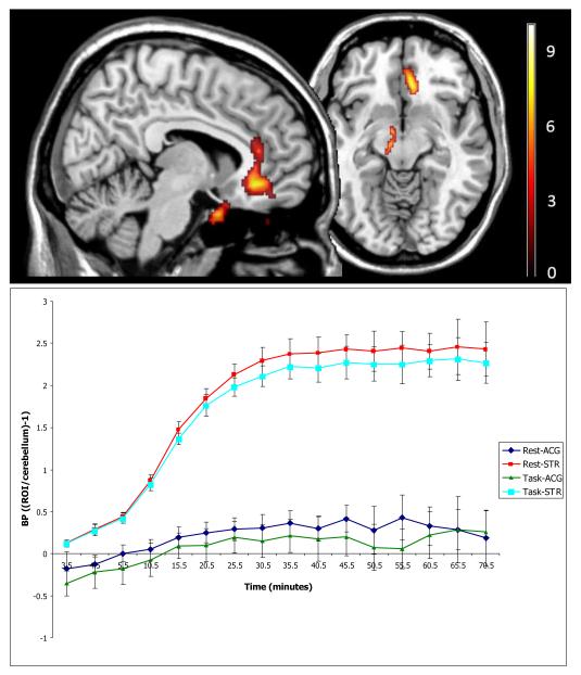
This systematic review describes human molecular imaging studies which have investigated alterations in extracellular DA levels during performance of behavioral tasks. Whilst heterogeneity in experimental methods limits meta-analysis, we describe the advantages and limitations of different methodological approaches. Interpretation of experimental results may be limited by regional cerebral blood flow (rCBF) changes, head movement and choice of control conditions. We revisit our original study of striatal DA release during video-game playing [Koepp, M.J., Gunn, R.N., Lawrence, A.D., Cunningham, V.J., Dagher, A., Jones, T., Brooks, D.J., Bench, C.J., Grasby, P.M., 1998. Evidence for striatal dopamine release during a video game. Nature 393, 266-268] to illustrate the potentially confounding influences of head movement and alterations in rCBF. Changes in [(11)C]raclopride binding may be detected in extrastriatal as well as striatal brain regions-however we review evidence which suggests that extrastriatal changes may not be clearly interpreted in terms of DA release. Whilst several investigations have detected increases in striatal extracellular DA concentrations during task components such as motor learning and execution, reward-related processes, stress and cognitive performance, the presence of potentially biasing factors should be carefully considered (and, where possible, accounted for) when designing and interpreting future studies.


Figures

Figure 1
Figure 1. Regional cerebral blood flow during performance of a videogame
Four healthy male volunteers each underwent a 12 run quantitative H2-[15]0 PET study. The volunteers were at rest during runs 1-5 and 11-12 and were playing a videogame during runs 6-10. A presents rCBF values for the dorsal and ventral striatum and cerebellum. The videogame was associated with increases in rCBF in all ROI, but to the greatest extent in the cerebellum. In B, data is presented as rCBF values in dorsal and ventral striatal ROI divided by rCBF values obtained in the cerebellum to provide ‘R1’ values. Here, decreases in ‘R1’ are apparent in striatal ROIs during the videogame.
Figure 2
Figure 2. Occupancy of the D2/3 receptors in different brain regions following administration of 400mg sulpiride
Seven healthy male volunteers (28-54 years, 65-85 kg) were administered either placebo or 400 mg of sulpiride. The subjects were dosed 90 min prior to the bolus injection of [11C]raclopride followed by a continuous infusion (Kbol=105 min, 170-340 MBq at start of scan). The 80-minute PET studies were performed on a CTI ECAT EXACT 3D tomograph offering a spatial resolution of 4.8 mm × 4.8 mm × 5.6 mm and an axial FOV of 23.4 cm. The dynamic PET images were realigned using FBF realignment after wavelet denoising. Each dynamic PET scan was summed for the equilibrium period (35 to 80 minutes) and was normalised to a [11C]raclopride PET template in MNI space. A probabilistic atlas (Hammers et al., 2003), also in MNI space, was placed over each normalised integrated PET images to generate the radioactivity concentration at equilibrium in the reference (Creference) and target (Ctarget) regions. The regional BP values were estimated using, BP = ((Ctarget − Creference)/Creference)) for both the placebo and sulpiride conditions. The regional occupancy values of the D2/3 receptors by sulpiride were calculated from the BP values as Occupancy = ((BPplacebo − BPdrug)/BPplacebo) × 100. Data is presented for seven subjects (shown as different coloured symbols) as the mean occupancy per region for all subjects (± standard deviation). Using a one-tailed paired t-test, the drug BP values were statistically lower from the placebo BP values in the striatal (p<0.001) and thalamic (p<0.02) regions, but not cortical areas (p>0.05). Due to the high degree of variance across subjects in cortical BP values, the mean occupancy in the cortical areas could not be estimated reliably In addition, some subjects showed negative occupancy in extrastriatal areas. This data has previously been presented in abstract form (Pretorius et al., 2004).
Figure 3
Figure 3. Scatterplot of extrastriatal binding potentials measured using two different dopamine D2 radiotracers ([11C]-raclopride and [11C]-FLB457) in the same 10 volunteers from Ito et al., (2008)
The plot was obtained using data presented in Ito et al. (2008). Correlational analysis shows that there is no significant relationship between ([11C]-raclopride and [11C]-FLB457 extrastriatal binding potentials (rs=0.032; p=0.92).
Figure 4
Figure 4. Change in extrastriatal [11C]-raclopride BP during a Tower of London planning task
A: BP values are presented in the planning task compared to rest, overlaid on a T1-weighted MRI image, analysed with SPM5. All clusters are significant at p<0.05 after correction for multiple comparisons across the entire volume, with a voxel height threshold of p<0.001. Sagittal section is at x=8, transverse section at y=−10. Statistically significant changes can be seen in the anterior cingulate cortex (ACG), in the region of the substantia nigra (left) and possibly the pituitary gland. B: An ROI was extracted around the significant cluster in the ACG and used to extract time-activity curves from individual scan data. Data is presented as mean ± standard deviation and the striatum (STR) is included for comparison. During the equilibrium sampling period (35-75 minutes), BP was significantly lower in the task condition in both the STR (p<0.05) and ACG (p<0.01). However, in the ACG but not the STR a separation in signal is apparent across the entire experiment. In addition, a high level of noise was associated with ACG BP measures. Experimental details are provided in Lappin et al., (2008).

References

    1. Aalto S, Bruck A, Laine M, Nagren K, Rinne JO. Frontal and temporal dopamine release during working memory and attention tasks in healthy humans: a positron emission tomography study using the high-affinity dopamine D2 receptor ligand [11C]FLB 457. J.Neurosci. 2005;25:2471–2477. - PMC - PubMed
    1. Abercrombie ED, Keefe KA, DiFrischia DS, Zigmond MJ. Differential effect of stress on in vivo dopamine release in striatum, nucleus accumbens, and medial frontal cortex. J.Neurochem. 1989;52:1655–1658. - PubMed
    1. Abi-Dargham A, Gil R, Krystal J, Baldwin RM, Seibyl JP, Bowers M, van Dyck CH, Charney DS, Innis RB, Laruelle M. Increased striatal dopamine transmission in schizophrenia: confirmation in a second cohort. Am.J.Psychiatry. 1998;155:761–767. - PubMed
    1. Alexander GE, DeLong MR, Strick PL. Parallel organization of functionally segregated circuits linking basal ganglia and cortex. Annu.Rev.Neurosci. 1986;9:357–381. - PubMed
    1. Alpert NM, Badgaiyan RD, Livni E, Fischman AJ. A novel method for noninvasive detection of neuromodulatory changes in specific neurotransmitter systems. Neuroimage. 2003;19:1049–1060. - PubMed

Publication types

MeSH terms