Review
. 2009 Aug;143(2):147-61.
doi: 10.1016/j.virusres.2009.05.010. Epub 2009 May 27.

Influenza virus morphogenesis and budding

Affiliations
Review

Influenza virus morphogenesis and budding

Debi P Nayak et al. Virus Res. 2009 Aug.

Abstract

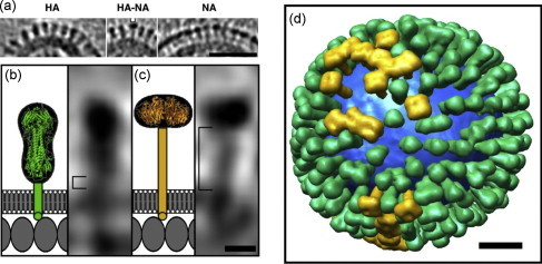
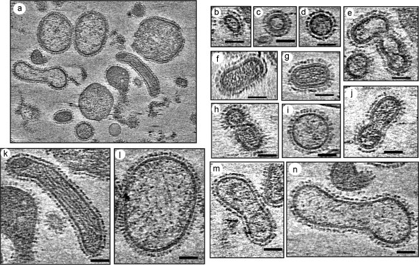
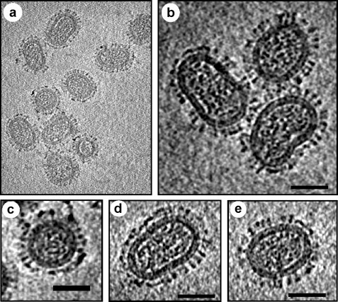
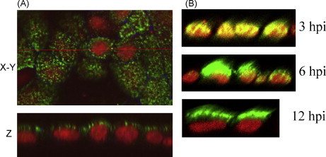
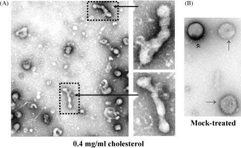
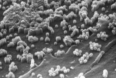
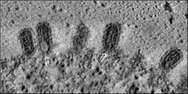
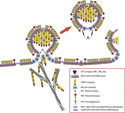
Influenza viruses are enveloped, negative stranded, segmented RNA viruses belonging to Orthomyxoviridae family. Each virion consists of three major sub-viral components, namely (i) a viral envelope decorated with three transmembrane proteins hemagglutinin (HA), neuraminidase (NA) and M2, (ii) an intermediate layer of matrix protein (M1), and (iii) an innermost helical viral ribonucleocapsid [vRNP] core formed by nucleoprotein (NP) and negative strand viral RNA (vRNA). Since complete virus particles are not found inside the cell, the processes of assembly, morphogenesis, budding and release of progeny virus particles at the plasma membrane of the infected cells are critically important for the production of infectious virions and pathogenesis of influenza viruses as well. Morphogenesis and budding require that all virus components must be brought to the budding site which is the apical plasma membrane in polarized epithelial cells whether in vitro cultured cells or in vivo infected animals. HA and NA forming the outer spikes on the viral envelope possess apical sorting signals and use exocytic pathways and lipid rafts for cell surface transport and apical sorting. NP also has apical determinant(s) and is probably transported to the apical budding site similarly via lipid rafts and/or through cortical actin microfilaments. M1 binds the NP and the exposed RNAs of vRNPs, as well as to the cytoplasmic tails (CT) and transmembrane (TM) domains of HA, NA and M2, and is likely brought to the budding site on the piggy-back of vRNP and transmembrane proteins. Budding processes involve bud initiation, bud growth and bud release. The presence of lipid rafts and assembly of viral components at the budding site can cause asymmetry of lipid bilayers and outward membrane bending leading to bud initiation and bud growth. Bud release requires fusion of the apposing viral and cellular membranes and scission of the virus buds from the infected cellular membrane. The processes involved in bud initiation, bud growth and bud scission/release require involvement both viral and host components and can affect bud closing and virus release in both positive and negative ways. Among the viral components, M1, M2 and NA play important roles in bud release and M1, M2 and NA mutations all affect the morphology of buds and released viruses. Disassembly of host cortical actin microfilaments at the pinching-off site appears to facilitate bud fission and release. Bud scission is energy dependent and only a small fraction of virus buds present on the cell surface is released. Discontinuity of M1 layer underneath the lipid bilayer, absence of outer membrane spikes, absence of lipid rafts in the lipid bilayer, as well as possible presence of M2 and disassembly of cortical actin microfilaments at the pinching-off site appear to facilitate bud fission and bud release. We provide our current understanding of these important processes leading to the production of infectious influenza virus particles.


Figures

Fig. 1
Fig. 1
Model virus with HA and NA spikes by cryoET analysis of X31 virus. (a) HA cluster (left); single NA (marked) in a cluster of HA (middle); cluster of mainly NA (right). (b and c) The stem length of HA and NA (square brackets in b and c, respectively. The structures of the stem, transmembrane domain and ectodomain are shown schematically. Molecules in the matrix layer are inferred to be packed in a monolayer (scale bar 5 nm). (d) Model of distribution of HA (green), NA (gold), and lipid bilayers (blue) in a single virion (scale 20 nm). Reproduced from Harris et al. (2006) with permission. The figure was provided by Drs A. Harris and A.C. Steven. (For interpretation of the references to color in this figure legend, the reader is referred to the web version of the article.)
Fig. 2
Fig. 2
Cryo-electron tomography studies of influenza virus A/PR8 strain showing highly pleomorphic virion architecture. (a) A density slice from a 3D cryo-electron tomography reconstruction of influenza A virus strain PR8. PR8 virus was grown in MDCK cells at 0.001 moi. The tilt series spanning −70° to 70° sample tilt were recorded in a TF20 cryo-electron microscope using the Batch Tomography program (FEI Company), and reconstructed using the Inspect3D (FEI Company) and refined by Protomo program (Winkler and Taylor, 2006). (b–n) Comparison of central slices of viral particles extracted from different cryo-electron tomograms. Different virus particles were picked at random. No attempt was made to determine the percentage of each virus form in the population. Each virus particle contained electron dense spots (RNP) inside, and spikes outside. Both HA and NA spikes, as identified based on morphology as described in Harris et al. (2006), were visible on the outer membrane. Scale bar 50 nm.
Fig. 3
Fig. 3
CryoET reconstruction of influenza virus A/WSN strain showing more homogeneous architecture as compared to PR8. The WSN strain virus particles were grown in MDCK cells at 0.05 moi. (a) A density slice from a 3D cryo-electron tomography reconstruction of influenza A strain WSN. Virus morphology was relatively less pleomorphic as compared to PR8 virus. (b–e) Comparison of central slices of viral particles extracted from the tomogram. Different virus particles were picked at random. No attempt was made to determine the percentage of each virus form in the population. Each virus particle contained electron dense spots (RNP) inside, and spikes outside. Both HA and NA spikes, as identified based on morphology as described in Harris et al. (2006), were visible on the outer membrane. Bar scale 50 nm.
Fig. 4
Fig. 4
Viral glycoprotein-independent apical transport of NP in polarized MDCK cells. VLP (delHA, del-NA, and del-M2-CT-TMD) infected (A) or wt virus-infected (B) MDCK cells were fixed (at 12 hpi. for panel A) with paraformaldehyde, permeabilized with saponin and triple-stained for NP (green), tight junction protein ZO-1 (blue) and DNA (red, with propidium iodide) and examined by confocal microscopy. Single optical section in the xy plane (A, upper, top/apical surface) and xz planes (A, lower and B) are shown for merged fluorescence. (For interpretation of the references to color in this figure legend, the reader is referred to the web version of the article.)
Fig. 5
Fig. 5
Deformed particles are produced in the presence of exogenous cholesterol. MDCK cells were infected (3.0 moi) with WSN virus and at 5 hpi virus-infected cells were either treated with 0.4 mg/ml water soluble cholesterol containing 6 mM MβCD (A) or mock treated (B). At 12 hpi, released virus particles were purified and examined by negative-stain EM (Barman and Nayak, 2007). Panel A, left picture was taken at 29,000×, and portions were further magnified at 72,000× (panel A, right). Panel B picture was taken at 72,000× magnification. Daisy-chain structures of some virus particles (panel A) released from cells in the presence of exogenous cholesterol indicate defective bud pinching-off process.
Fig. 6
Fig. 6
Scanning electron micrographs of spheroidal influenza virus buds attached to infected cells (40,000×). This micrograph was kindly provided by and is printed with the permission of David Hockley of the National Institute of Biological Standard and Control at Hertfordshire, UK.
Fig. 7
Fig. 7
Virus buds at the cell surface by ET. At 12 hpi WSN-infected MDCK cells were processed for thin section and examined by ET. This picture represents one slice through the central region of the virus buds. One can see the parallel arrangement of the vRNPs inside the bud perpendicular to cell surface. The bud neck (⇒) shows gaps indicating possible absence of M1. HA and NA spikes are seen on the bud envelope.
Fig. 8
Fig. 8
Schematic illustration of the pinching-off process of influenza virus bud. The pinching-off region (neck) is shown to be viral membrane devoid of lipid rafts (Barman and Nayak, 2007), devoid of HA and NA spikes outside and M1 inside the lipid bilayers (Harris et al., 2006) and may contain M2 (Schroeder et al., 2005).

References

    1. Akarsu H., Burmeister W.P., Petosa C., Petit I., Muller C.W., Ruigrok R.W., Baudin F. Crystal structure of the M1 protein-binding domain of the influenza A virus nuclear export protein (NEP/NS2) EMBO J. 2003;22(18):4646–4655. - PMC - PubMed
    1. Ali A., Avalos R.T., Ponimaskin E., Nayak D.P. Influenza virus assembly: effect of influenza virus glycoproteins on the membrane association of M1 protein. J. Virol. 2000;74(18):8709–8719. - PMC - PubMed
    1. Avalos R.T., Yu Z., Nayak D.P. Association of influenza virus NP and M1 proteins with cellular cytoskeletal elements in influenza virus-infected cells. J. Virol. 1997;71(4):2947–2958. - PMC - PubMed
    1. Bancroft C.T., Parslow T.G. Evidence for segment-nonspecific packaging of the influenza a virus genome. J. Virol. 2002;76(14):7133–7139. - PMC - PubMed
    1. Barman S., Adhikary L., Chakraborti A.K., Bernas C., Kawaoka Y., Nayak D.P. Role of transmembrane domain and cytoplasmic tail amino acid sequences of influenza A virus neuraminidase in raft-association and virus budding. J. Virol. 2004;78(10):5258–5269. - PMC - PubMed

Publication types