

Reducing inter-subject anatomical variation: effect of normalization method on sensitivity of functional magnetic resonance imaging data analysis in auditory cortex and the superior temporal region
- PMID: 19481162
- PMCID: PMC2804864
- DOI: 10.1016/j.neuroimage.2009.05.047
Reducing inter-subject anatomical variation: effect of normalization method on sensitivity of functional magnetic resonance imaging data analysis in auditory cortex and the superior temporal region
Abstract
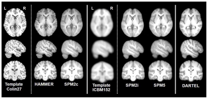
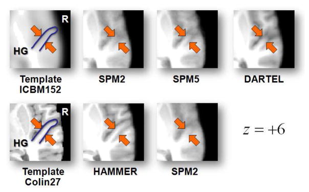
Conventional group analysis of functional MRI (fMRI) data usually involves spatial alignment of anatomy across participants by registering every brain image to an anatomical reference image. Due to the high degree of inter-subject anatomical variability, a low-resolution average anatomical model is typically used as the target template, and/or smoothing kernels are applied to the fMRI data to increase the overlap among subjects' image data. However, such smoothing can make it difficult to resolve small regions such as subregions of auditory cortex when anatomical morphology varies among subjects. Here, we use data from an auditory fMRI study to show that using a high-dimensional registration technique (HAMMER) results in an enhanced functional signal-to-noise ratio (fSNR) for functional data analysis within auditory regions, with more localized activation patterns. The technique is validated against DARTEL, a high-dimensional diffeomorphic registration, as well as against commonly used low-dimensional normalization techniques such as the techniques provided with SPM2 (cosine basis functions) and SPM5 (unified segmentation) software packages. We also systematically examine how spatial resolution of the template image and spatial smoothing of the functional data affect the results. Only the high-dimensional technique (HAMMER) appears to be able to capitalize on the excellent anatomical resolution of a single-subject reference template, and, as expected, smoothing increased fSNR, but at the cost of spatial resolution. In general, results demonstrate significant improvement in fSNR using HAMMER compared to analysis after normalization using DARTEL, or conventional normalization such as cosine basis function and unified segmentation in SPM, with more precisely localized activation foci, at least for activation in the region of auditory cortex.
Figures
References
-
- Ardekani B, Guckemus S, Bachman A, Hoptman M, Wojtaszek M, Nierenberg J. Quantitative comparison of algorithms for inter-subject registration of 3D volumetric brain MRI scans. Journal of Neuroscience Methods. 2005;142:67–76. - PubMed
-
- Ardekani BA. An improved method for intersubject registration in 3D volumetric brain MRI. Sydney, Australia. World Congress on Medical Physics and Biomedical Engineering; 2003. Abstract 452.
-
- Ardekani BA, Bachman AH, Strother S, Fujibayashi Y, Yonekura Y. Impact of inter-subject image registration on group analysis of fMRI data. International Congress Series, Elsevier. 2004;1265:49–59.
-
- Ashburner J. A fast diffeomorphic image registration algorithm. NeuroImage. 2007;38:95–113. - PubMed
-
- Ashburner J, Friston K. Fully three-dimensional nonlinear spatial normalization: A new approach. NeuroImage. 1996;3:S111.
Publication types
MeSH terms
Grants and funding
LinkOut - more resources
Full Text Sources
Medical
