. 2009 Jun;84(6):771-9.
doi: 10.1016/j.ajhg.2009.05.003. Epub 2009 May 28.

X chromosome inactivation is initiated in human preimplantation embryos

Affiliations

X chromosome inactivation is initiated in human preimplantation embryos

Ilse M van den Berg et al. Am J Hum Genet. 2009 Jun.

Abstract

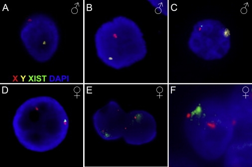
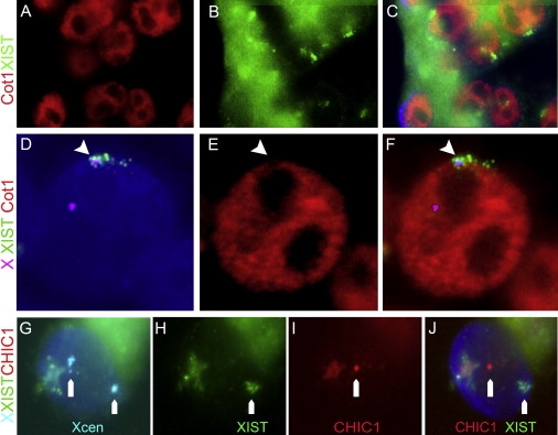
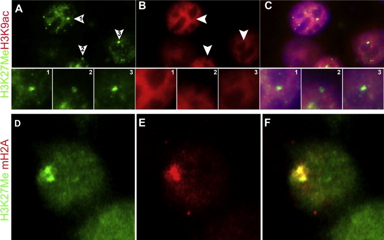
X chromosome inactivation (XCI) is the mammalian mechanism that compensates for the difference in gene dosage between XX females and XY males. Genetic and epigenetic regulatory mechanisms induce transcriptional silencing of one X chromosome in female cells. In mouse embryos, XCI is initiated at the preimplantation stage following early whole-genome activation. It is widely thought that human embryos do not employ XCI prior to implantation. Here, we show that female preimplantation embryos have a progressive accumulation of XIST RNA on one of the two X chromosomes, starting around the 8-cell stage. XIST RNA accumulates at the morula and blastocyst stages and is associated with transcriptional silencing of the XIST-coated chromosomal region. These findings indicate that XCI is initiated in female human preimplantation-stage embryos and suggest that preimplantation dosage compensation is evolutionarily conserved in placental mammals.


Figures

Figure 1
Figure 1
XIST Expression in Male and Female Human Preimplantation Embryos RNA and DNA FISH staining with probes detecting XIST RNA (green), the X (red) and Y (yellow) chromosome, and DAPI counterstain. Human male embryos (A–C) do not generally show XIST signals at the 8-cell stage (A) or at the morula stage (B). A minority of male cells at the morula stage show a pinpoint of XIST staining (C). Female embryos (D–F) show an XIST pinpoint in the majority of embryos at the 8-cell stage (D). Two cells at the morula stage each show a beginning cloud of XIST on one of their two X chromosomes (E). At the blastocyst stage, this has further accumulated to a full cloud on one of the two X chromosomes (F). A third diffuse red signal is an X chromosome from an adjacent cell that is in a different focal plane.
Figure 2
Figure 2
Transcriptional Changes on the Inactive X Chromosome (A–F) Cot1 exclusion around XIST. (A–C) Cells of a female blastocyst embryo, stained for Cot1 RNA (red in A) and XIST RNA (green in B) showing depleted regions of Cot-1 RNA around the XIST signals indicating the position of the Xi chromosome (merged in C). (D–F) Representative cell of a female blastocyst with staining for the X centromeres and XIST RNA (D; Xcen in magenta, XIST in green) together with Cot1 (red in E). Transcription of Cot1 RNA was absent in a region that overlaps with XIST RNA staining (F), whereas the active X without XIST staining overlaps with a Cot1-positive region. (G–J) Female blastocyst cell with two X centromeres (cyan in G) has a single XIST cloud on one X chromosome (green in H) and monoallelic expression of CHIC1 on the other X chromosome (red in I, merged in J). A dust spot is visible in all colors and is therefore nonspecific staining.
Figure 3
Figure 3
Epigenetic Changes on the Inactive X Chromosome (A–C) Three adjacent blastocyst cells show a single nuclear domain with H3K27Me3 hypermethylation (arrowheads in J and enlarged panels 1–3), and staining for H3K9 acetylation (B, 1–3) shows an H3K9ac-depleted region overlaying the H3K27Me3 accumulation (C, 1–3), indicating the position of the Xi chromosome. (D–F) Representative blastocyst cell shows a single nuclear domain with H3K27 hypermethylation (green, D) and enrichment for macroH2A (red, E) with a clear overlap (yellow, F), analogous to the signal around an Xi chromosome.

References

    1. Brockdorff N., Ashworth A., Kay G.F., Cooper P., Smith S., McCabe V.M., Norris D.P., Penny G.D., Patel D., Rastan S. Conservation of position and exclusive expression of mouse Xist from the inactive X chromosome. Nature. 1991;351:329–331. - PubMed
    1. Brown C.J., Ballabio A., Rupert J.L., Lafreniere R.G., Grompe M., Tonlorenzi R., Willard H.F. A gene from the region of the human X inactivation centre is expressed exclusively from the inactive X chromosome. Nature. 1991;349:38–44. - PubMed
    1. Penny G.D., Kay G.F., Sheardown S.A., Rastan S., Brockdorff N. Requirement for Xist in X chromosome inactivation. Nature. 1996;379:131–137. - PubMed
    1. Heard E., Disteche C.M. Dosage compensation in mammals: Fine-tuning the expression of the X chromosome. Genes Dev. 2006;20:1848–1867. - PubMed
    1. Lyon M.F. Gene action in the X-chromosome of the mouse (Mus musculus L.) Nature. 1961;190:372–373. - PubMed

Publication types