

Functional MRI reveals compromised neural integrity of the face processing network in congenital prosopagnosia
- PMID: 19481456
- PMCID: PMC2711224
- DOI: 10.1016/j.cub.2009.04.060
Functional MRI reveals compromised neural integrity of the face processing network in congenital prosopagnosia
Abstract
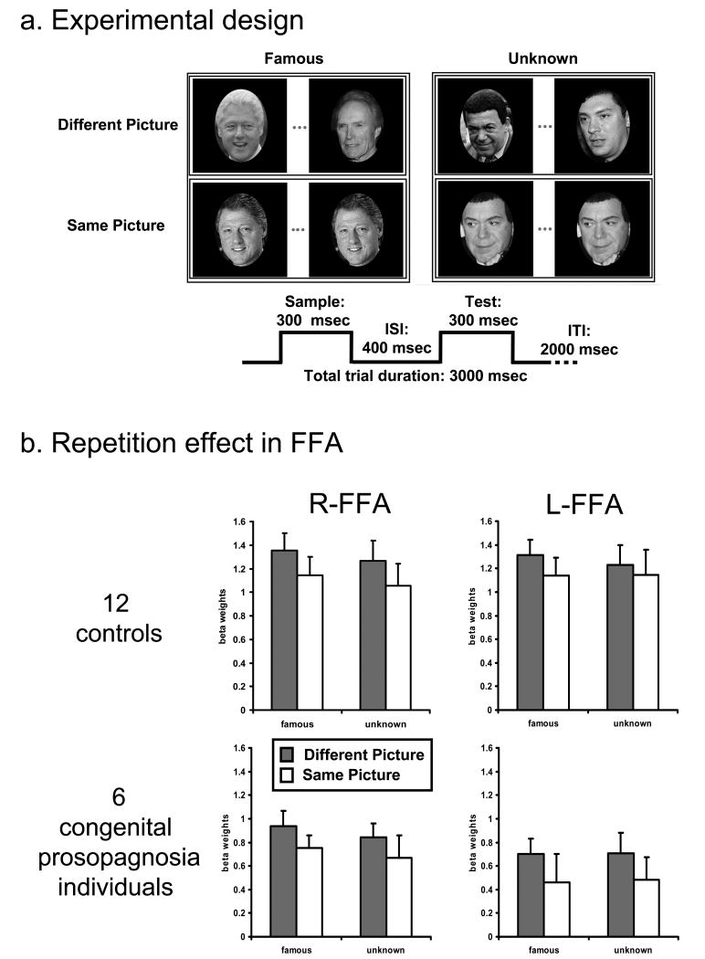
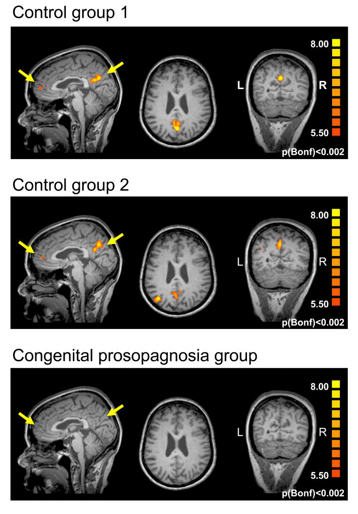
The summed activity of multiple nodes of a distributed cortical network supports face recognition in humans, including "core" ventral occipitotemporal cortex (VOTC) regions, and "extended" regions outside VOTC. Many individuals with congenital prosopagnosia-an impairment in face processing-exhibit normal blood oxygenation level-dependent (BOLD) activation in the core VOTC regions. These individuals evince a reduction in the structural integrity of the white matter tracts connecting VOTC to anterior temporal and frontal cortices, part of the "extended" face network. The impairment in congenital prosopagnosia may arise, therefore, not from a dysfunction of the core VOTC areas but from a failure to propagate signals between the intact VOTC and the extended nodes of the network. Using the fMR adaptation paradigm with famous and unknown faces, we show that individuals with congenital prosopagnosia evince normal adaptation effects in VOTC, indicating sensitivity to facial identity, but show no differential activation for familiar versus unknown faces outside VOTC, particularly in the precuneus/posterior cingulate cortex and the anterior paracingulate cortex. Normal BOLD activation in VOTC is thus insufficient to subserve intact face recognition, and disrupted information propagation between VOTC and the extended face processing network may explain the functional impairment in congenital prosopagnosia.
Figures
References
-
- Barton JJ. Disorders of face perception and recognition. Neurol Clin. 2003;21:521–548. - PubMed
-
- Rossion B, Caldara R, Seghier M, Schuller AM, Lazeyras F, Mayer E. A network of occipito-temporal face-sensitive areas besides the right middle fusiform gyrus is necessary for normal face processing. Brain. 2003;126:2381–2395. - PubMed
-
- Pitcher D, Walsh V, Yovel G, Duchaine B. TMS evidence for the involvement of the right occipital face area in early face processing. Curr Biol. 2007;17:1568–1573. - PubMed
-
- Haxby JV, Hoffman EA, Gobbini MI. The distributed human neural system for face perception. Trends Cogn Sci. 2000;4:223–233. - PubMed
-
- Ishai A. Let's face it: it's a cortical network. Neuroimage. 2008;40:415–419. - PubMed
Publication types
MeSH terms
Grants and funding
LinkOut - more resources
Full Text Sources
Other Literature Sources
Medical
