

doi: 10.1016/j.stem.2009.05.005.
Epub 2009 May 28.
Generation of human induced pluripotent stem cells by direct delivery of reprogramming proteins
Affiliations
- PMID: 19481515
- PMCID: PMC2705327
- DOI: 10.1016/j.stem.2009.05.005
Item in Clipboard
Generation of human induced pluripotent stem cells by direct delivery of reprogramming proteins
Cell Stem Cell.
.
No abstract available
Figures
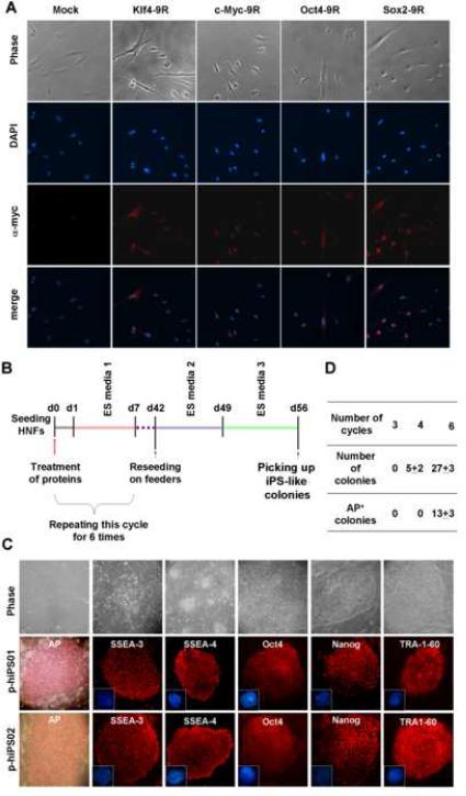
A, HNFs were incubated with HEK 293 extracts expressing each reprogramming protein and subjected to immunocytochemistry using myc antibodies. Nuclei were counterstained with DAPI. B, The schematic protocol depicts a repeated process and the time line for generating p-hiPS cells from HNFs. C, Top panel: starting HNFs (first image); morphology after 3 cycle protein treatments (second image); and increased colony number after 6 cycles (third image). Approximately half of these iPS-like colonies stained positive for AP; early morphology after p-hiPS colonies were transferred to MEF (fourth image); and morphology of established p-hiPS cell line at passage number 10 (p-hiPS01 [fifth image] and p-hiPS02 [sixth image]). Immunostaining of p-hiPS01 (middle panel) and p-hiPS02 (bottom panel) clones show expression of hESC markers, including AP, SSEA-3, SSEA-4, Oct-4, Nanog, and TRA-1-60. Nuclei were stained with DAPI (blue in second and third row panel). D, Efficiency of reprogrammed colony formation with iPS-like morphology and AP-positive staining after different numbers of the protein treatment cycle. This is the summary of three independent experiments with the standard error.
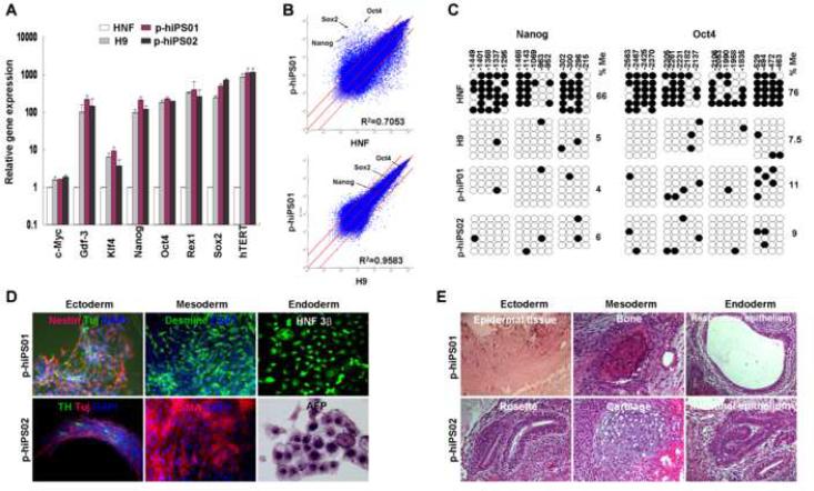
A, Quantitative RT-PCR was performed to assess the expression of c-Myc, Gdf-3, Klf4, Nanog, Oct4, Rex1, Sox2, and hTERT, in p-hiPS01 and p-hiPS02, hES (H9), and HNF cells. Relative gene expression represents fold changes relative to that of HNF cells normalized to β-actin expression. This experiment (repeated twice in triplicate using independently prepared cDNAs) resulted in almost identical patterns. B, The global gene-expression patterns were compared between p-hiPS01 and HNF, and between p-hiPS01 and H9 with Affymetrix microarrays. The red lines indicate the diagonal and 5-fold changes between the paired samples. C, Bisulfite sequencing analysis of the Nanog and Oct4 promoters reveal that almost complete epigenetic reprogramming. Open and closed circles indicate unmethylated and methylated CpG, respectively. Numbers on top show each CpG location. Percentages of CpG methylation (%Me) are shown. D. In vitro differentiation of p-hiPS cells. EBs were made by suspension culture of both p-hiPS lines at day 8 (Far left of the top row panels). Phase contrast (Top row panels) and immunostaining images (second and third row panels) show all three germ layer cells at day 24 including neural (ectodermal), muscle and endothelial-like (mesodermal), and endoderm-like cells (endoderm). E, Teratoma formation in immunodeficiency mice by p-iPS cells. H&E staining were performed for teratomas. The resulting teratomas contained tissues representing all three germ layers: (p-hiPS01, fourth row; and p-hiPS02, bottom row). Ectoderm: epidermal and neural tissue (rosette); mesoderm: bone and cartilage; endoderm: respiratory epithelium and intestinal-like epithelium.
References
-
- Belting M, Sandgren S, Wittrup A. Nuclear delivery of macromolecules: barriers and carriers. Adv Drug Deliv Rev. 2005;57:505–527. - PubMed
-
- Chung Y, Bishop CE, Treff NR, Walker SJ, Sandler VM, Becker S, Klimanskaya I, Wun WS, Dunn R, Hall RM, et al. Reprogramming of Human Somatic Cells Using Human and Animal Oocytes. Cloning Stem Cells. 2009 - PubMed
-
- Frankel AD, Bredt DS, Pabo CO. Tat protein from human immunodeficiency virus forms a metal-linked dimer. Science. 1988;240:70–73. - PubMed
-
- Frankel AD, Pabo CO. Cellular uptake of the tat protein from human immunodeficiency virus. Cell. 1988;55:1189–1193. - PubMed
Publication types
MeSH terms
Substances
Associated data
- Actions
- Actions
- Actions
- Actions
- Actions
- Actions
Grants and funding
LinkOut - more resources
Full Text Sources
Other Literature Sources
Molecular Biology Databases
Research Materials
