. 2009 May 14;34(4):440-50.
doi: 10.1016/j.molcel.2009.04.017.

A convergence of rRNA and mRNA quality control pathways revealed by mechanistic analysis of nonfunctional rRNA decay

Affiliations

A convergence of rRNA and mRNA quality control pathways revealed by mechanistic analysis of nonfunctional rRNA decay

Sarah E Cole et al. Mol Cell. .

Abstract

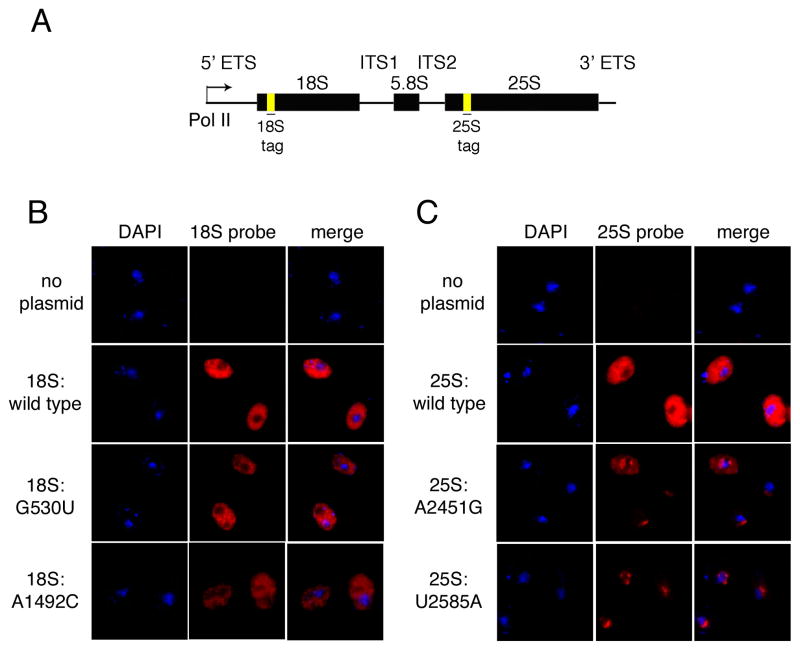
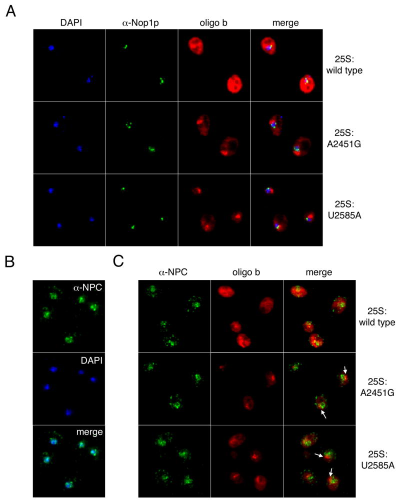
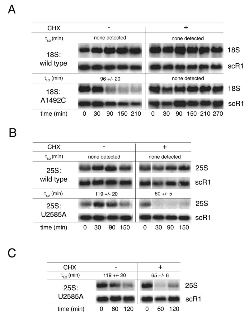
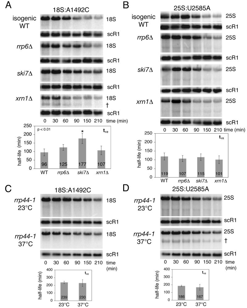
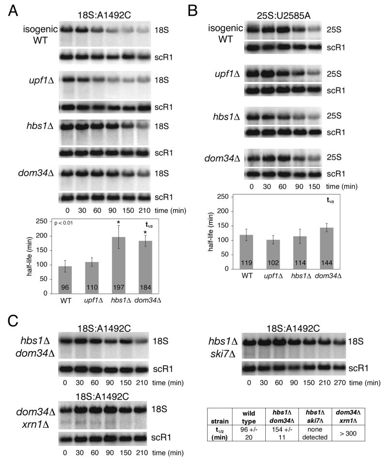
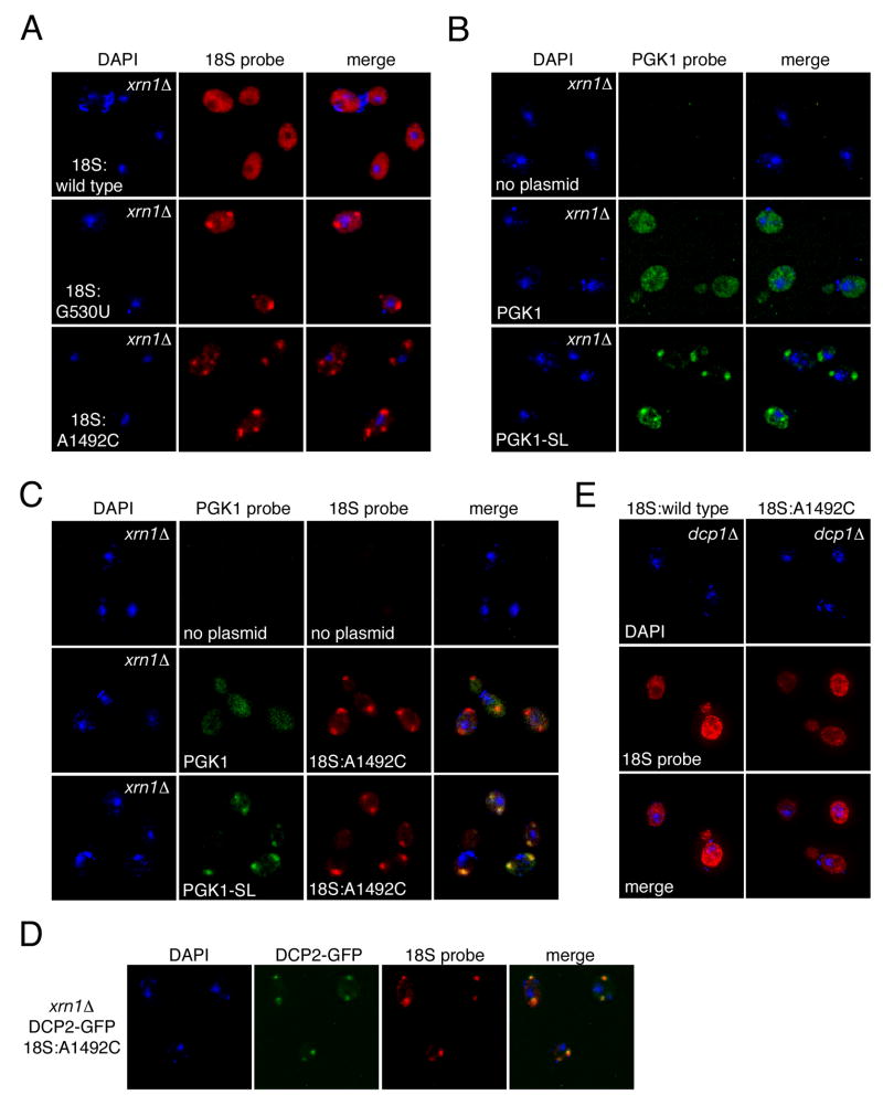
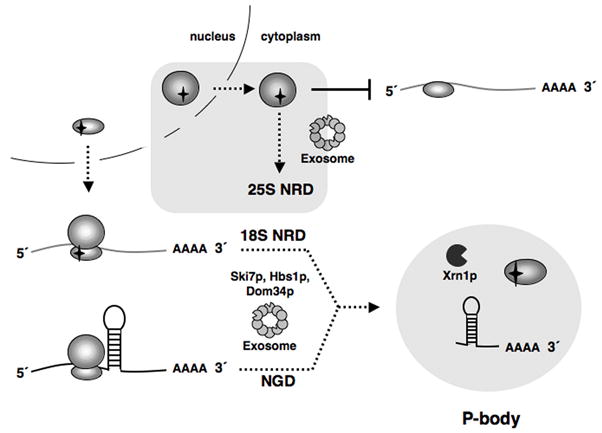
Eukaryotes possess numerous quality control systems that monitor both the synthesis of RNA and the integrity of the finished products. We previously demonstrated that Saccharomyces cerevisiae possesses a quality control mechanism, nonfunctional rRNA decay (NRD), capable of detecting and eliminating translationally defective rRNAs. Here we show that NRD can be divided into two mechanistically distinct pathways: one that eliminates rRNAs with deleterious mutations in the decoding site (18S NRD) and one that eliminates rRNAs containing deleterious mutations in the peptidyl transferase center (25S NRD). 18S NRD is dependent on translation elongation and utilizes the same proteins as those participating in no-go mRNA decay (NGD). In cells that accumulate 18S NRD and NGD decay intermediates, both RNA types can be seen in P-bodies. We propose that 18S NRD and NGD are different observable outcomes of the same initiating event: a ribosome stalled inappropriately at a sense codon during translation elongation.


Figures

Figure 1
Figure 1
In situ localization of wild type and mutant rRNAs. (A) Schematic of rDNA reporter plasmids containing neutral sequence tags at indicated sites. (B) FISH analysis of wild type cells (BY4741) carrying no plasmid, pJV12-WT (18S:wild type), pJV12-G530U (18S:G530U) or pJV12-A1492C (18S:A1492C) using Alexa 594-labeled probe FL125. (C) FISH analysis of wild type cells (BY4741) carrying no plasmid, pJV12-WT (25S:wild type), pJV12-A2451G (25S:A2451G) or pJV12-U2585A (25S:U2585A) using Alexa 594-labeled probe FL126. In both B and C, the nucleus is marked by DAPI staining.
Figure 2
Figure 2
Comparison of 25S rRNA localization with nucleolar and nuclear pore complex markers. (A) Wild type (BY4741) cells carrying pJV12-WT (25S:wild type), pJV12-A2451G (25S:A2451G) or pJV12-U2585A (25S:U2585A) analyzed by FISH using Alexa 594-labeled probe FL126 and by indirect immunofluorescence using an antibody recognizing the nucleolar protein Nop1p plus an Alexa 488-conjugated secondary antibody. (B) Detection of the nuclear envelope by indirect immunofluorescence using an antibody against nuclear pore complex O-linked glycoproteins and an Alexa 488-conjugated secondary antibody. In both A and B, DAPI staining shows the location of the nucleus. (C) Wild type (BY4741) cells carrying pJV12-WT (25S:wild type), pJV12-A2451G (25S:A2451G) or pJV12-U2585A (25S:U2585A) were analyzed by immunofluorescence to visualize the nuclear envelope as in B and FISH to visualize 25S rRNAs as in A. Arrows indicate foci of mutant 25S rRNA outside the nucleus.
Figure 3
Figure 3
Differential effects of cycloheximide on 18S and 25S NRD. (A) Transcriptional pulse-chase and northern analysis of wild type cells (BY4741) carrying either pSC40 (18S:wild type) or pSC40-A1492C (18S:A1492C) in the absence or presence of cycloheximide. (B) Transcriptional pulse-chase and northern analysis of wild type cells (BY4741) carrying either pSC40 (25S:wild type) or pSC40-U2585A (25S:U2585A) in the absence or presence of cycloheximide. Cycloheximide was added at transcriptional shut off. (C) Same as in B except cycloheximide was added 30 minutes after transcriptional shut off. In both A and B, times indicated are relative to transcriptional shut off (i.e., glucose addition; t=0). In C, times indicated are relative to cycloheximide addition. In B and C cultures were split at the time of cycloheximide addition and thus share the t=0 time point. Plasmid-derived rRNAs were detected with 32P-labeled probe FL125 or FL126. Endogenous scR1 RNA, which served as a loading control, was also monitored by northern blotting. Indicated half-lives are the mean of at least three independent trials (error: standard deviation).
Figure 4
Figure 4
Analysis of 18S and 25S NRD in exonuclease-deficient strains. (A) Transcriptional pulse-chase and northern analysis of 18S:A1492C rRNA expressed from pSC40-A1492C in wild type strain BY4741 or the indicated isogenic exonuclease-deficient strains. indicates an 18S rRNA decay intermediate. (B) Same as A, except strains contained pSC40-U2585A expressing 25S:U2585A rRNA. (C) Transcriptional shut off and northern analysis of 18S:A1492C rRNA expressed from pSC40-A1492C in the temperature-sensitive rrp44-1 strain grown in galactose at the permissive temperature (23°C) or shifted to the non-permissive temperature (37°C) 3 hours prior to glucose addition. (D) Same as C, except strains contained pSC40-U2585A expressing 25S:U2585A rRNA. indicates a 25S rRNA decay intermediate. In all panels, times indicated are relative to transcriptional shut off (i.e., glucose addition; t=0). Plasmid-derived rRNAs and endogenous scR1 RNA, which served as a loading control, were detected with 32P-labeled probes (FL125, FL126 and FL217). Histograms report average rRNA half-lives from at least three independent trials (error: standard deviation). Mutant strains exhibiting statistically significant differences (unpaired student t-test; p<0.01) in 18S:A1492C rRNA half-lives are indicated (*).
Figure 5
Figure 5
Analysis of 18S and 25S NRD in strains lacking mRNA decay proteins. (A) Transcriptional pulse-chase analysis of 18S:A1492C rRNA expressed from pSC40-A1492C in wild type strain BY4741 or indicated isogenic mutant strains. (B) Same as A, except strains contained pSC40-U2585A expressing 25S:U2585A rRNA. (C) Same as A. Double mutant strains are isogenic to BY4741. In A and B, the isogenic wild type time courses shown are the same as those in Figure 4A and B; experiments in these panels were preformed concurrently. In all panels, times indicated are relative to transcriptional shut off (i.e., glucose addition; t=0). Plasmid-derived rRNAs and endogenous scR1 RNA, which served as a loading control, were detected with 32P-labeled probes (FL125, FL126 and FL217). Histograms report average rRNA half-lives from at least three independent trials (error: standard deviation). Mutant strains exhibiting statistically significant differences (unpaired student t-test; p<0.01) in 18S:A1492C rRNA half-lives are indicated (*).
Figure 6
Figure 6
FISH analysis of 18S NRD and NGD substrates. (A) xrn1Δ strain (YGL173C) carrying pJV12-WT (18S:wild type), pJV12-G530U (18S:G530U) or pJV12-A1492C (18S:A1492C) analyzed by FISH using Alexa 594-labeled probe FL125. (B) FISH analysis of xrn1Δ strain (HFY1081) carrying reporter plasmid PGK1 (pRP469) or PGK1-SL (pRP1251) using Alexa 647-labeled probe RP141. (C) xrn1Δ strain (HFY1081) carrying pWL160-A1492C and PGK1-SL (pRP1251) analyzed by FISH using Alexa 647-labeled probe RP141 and Cy3-labeled probe FL125. (D) FISH analysis of xrn1Δ DCP2-GFP strain (yRP1923) carrying pWL160-A1492C using Cy3-labeled probe FL125. (E) FISH analysis of dcp1Δ DHH1-GFP strain (yRP1736) carrying pSC39 or pWL160-A1492C using Alexa 594-labeled probe FL125.
Figure 7
Figure 7
Models for 18S NRD and 25S NRD. Upon export from the nucleus, 18S NRD substrates are able to engage in translation. However, deleterious decoding site mutations cause these defective 40S subunits to stall on mRNAs, a scenario similar to ribosome stalling on an NGD substrate due to a structural hindrance in the mRNA. In both cases, the stalled translation complexes are eliminated through the combined action of Dom34p, Hbs1p, Ski7p, Xrn1p and the cytoplasmic exosome. 5′->3′ decay of 18S NRD and NGD substrates occurs in P-bodies. Conversely, 25S NRD substrates, which accumulate around the nuclear envelope, are eliminated after export to the cytoplasm in a process not requiring ongoing translation elongation but involving the core exosome.

Comment in

Similar articles

Cited by

References

    1. Amberg DC, Goldstein AL, Cole CN. Isolation and characterization of RAT1: an essential gene of Saccharomyces cerevisiae required for the efficient nucleocytoplasmic trafficking of mRNA. Genes Dev. 1992;6:1173–1189. - PubMed
    1. Amrani N, Sachs MS, Jacobson A. Early nonsense: mRNA decay solves a translational problem. Nat Rev Mol Cell Biol. 2006;7:415–425. - PubMed
    1. Benard L, Carroll K, Valle RC, Masison DC, Wickner RB. The ski7 antiviral protein is an EF1-alpha homolog that blocks expression of non-Poly(A) mRNA in Saccharomyces cerevisiae. J Virol. 1999;73:2893–2900. - PMC - PubMed
    1. Bousquet-Antonelli C, Presutti C, Tollervey D. Identification of a regulated pathway for nuclear pre-mRNA turnover. Cell. 2000;102:765–775. - PubMed
    1. Brengues M, Parker R. Accumulation of polyadenylated mRNA, Pab1p, eIF4E, and eIF4G with P-bodies in Saccharomyces cerevisiae. Mol Biol Cell. 2007;18:2592–2602. - PMC - PubMed

Publication types

MeSH terms