. 2009 May 14;34(4):473-84.
doi: 10.1016/j.molcel.2009.04.021.

Structural insights into the regulatory particle of the proteasome from Methanocaldococcus jannaschii

Affiliations

Structural insights into the regulatory particle of the proteasome from Methanocaldococcus jannaschii

Fan Zhang et al. Mol Cell. .

Abstract

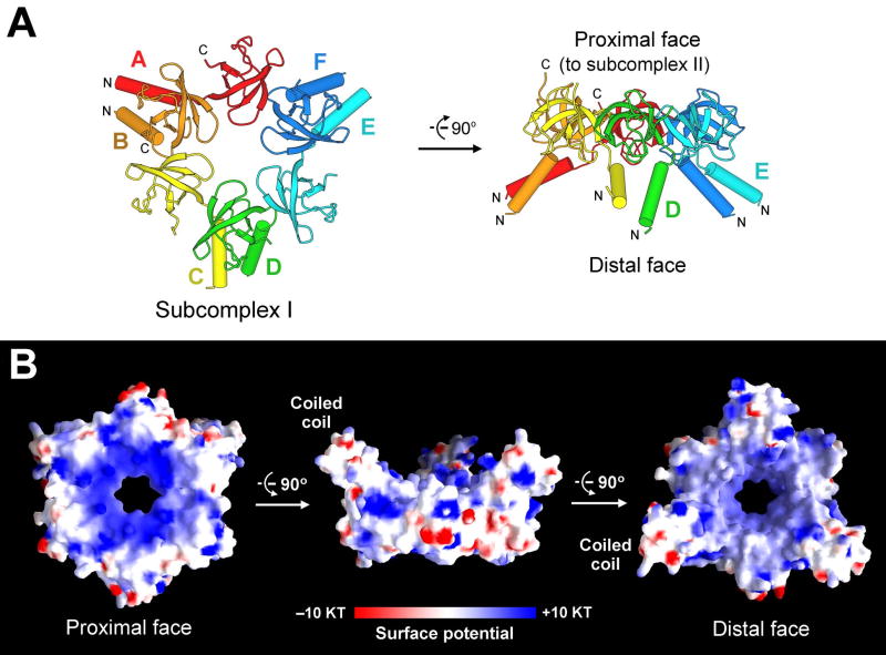
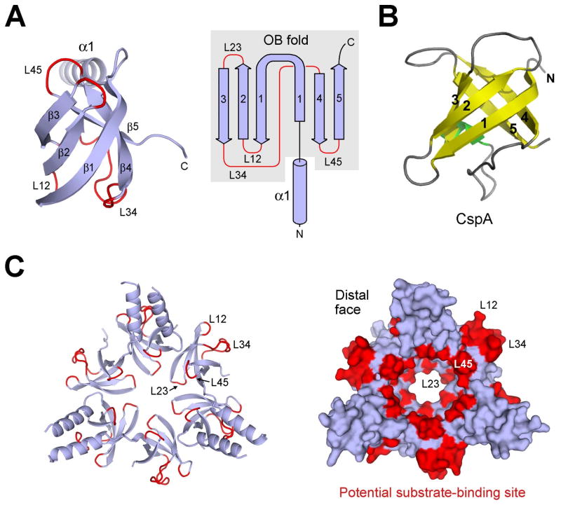
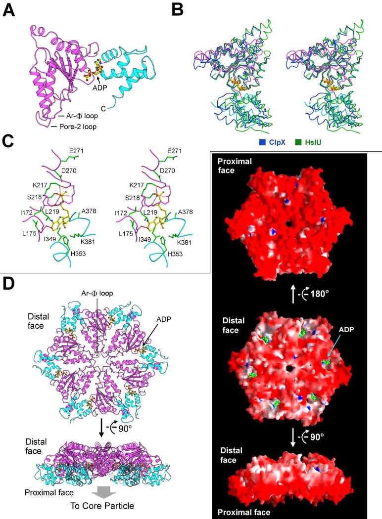
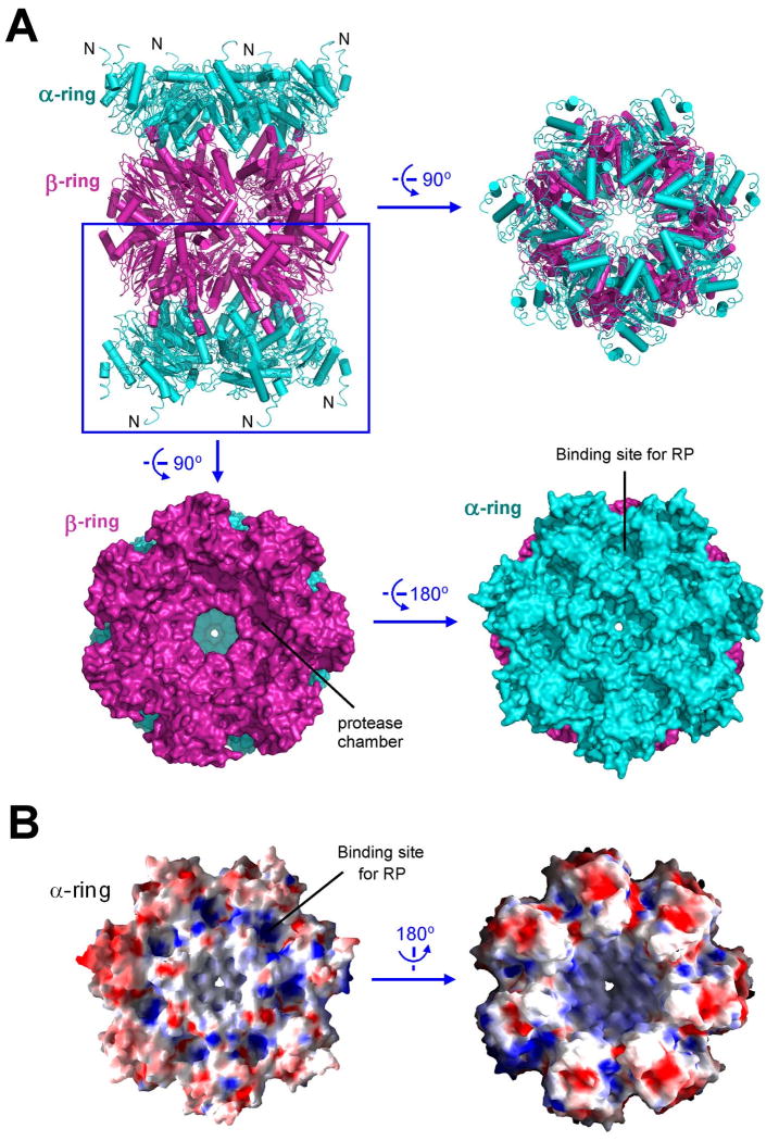
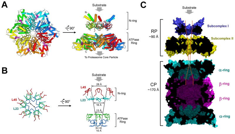
Eukaryotic proteasome consists of a core particle (CP), which degrades unfolded protein, and a regulatory particle (RP), which is responsible for recognition, ATP-dependent unfolding, and translocation of polyubiquitinated substrate protein. In the archaea Methanocaldococcus jannaschii, the RP is a homohexameric complex of proteasome-activating nucleotidase (PAN). Here, we report the crystal structures of essential elements of the archaeal proteasome: the CP, the ATPase domain of PAN, and a distal subcomplex that is likely the first to encounter substrate. The distal subcomplex contains a coiled-coil segment and an OB-fold domain, both of which appear to be conserved in the eukaryotic proteasome. The OB domains of PAN form a hexameric ring with a 13 A pore, which likely constitutes the outermost constriction of the substrate translocation channel. These studies reveal structural codes and architecture of the complete proteasome, identify potential substrate-binding sites, and uncover unexpected asymmetry in the RP of archaea and eukaryotes.


Figures

Figure 1
Figure 1
Structure of subcomplex I of the PAN regulatory particle. (A) Structure of the hexameric subcomplex I. The 6 Pan fragments are labeled A through F. Each Pan fragment (residues 74-150) contains an N-terminal α-helix followed by a β-domain that is structurally homologous to the oligonucleotide/oligosaccharide binding fold (OB-fold). Two α-helices from adjacent domains form a coiled coil. The distal and proximal faces refer to their relative orientation with respect to the 20S core particle. (B) Surface potential of subcomplex I. The positive and negative charges are colored blue and red, respectively.
Figure 2
Figure 2
Structure-based sequence alignment between PAN and the AAA+ ATPases in yeast and human proteasomes. Rpt1 through Rpt6 are derived from S. cerevisiae. Secondary structural elements of PAN are indicated above the sequences. Invariant and conserved amino acids are colored yellow and cyan, respectively. Residues that correspond to Pro91, Asp99, and Arg134 of PAN, are highlighted in red and green. Doubled arrows indicate residues that are predicted to line the substrate translocation channel. The loop that falls between β8 and α5 is known as pore loop 1 (pore-1 loop, or Ar-Φ loop), and that between α8 and α9 is pore loop 2 (pore-2 loop). The OB domain pore loops are termed L23 and L45.
Figure 3
Figure 3
The β-domain of PAN exhibits an oligonucleotide/oligosaccharide binding fold (OB-fold). (A) Ribbon (left panel) and topology (right panel) diagrams of the β-domain of PAN reveal an OB-fold. The surface loops connecting adjacent β-strands are highlighted in red. (B) Structure of a representative OB-fold protein CspA (Schindelin et al., 1994). (C) Candidate substrate-binding site in subcomplex I. Subcomplex I is represented in ribbon diagram (left panel) and surface potential (right panel). The surface loops are concentrated in two areas: inner surface of the central passage (L23 and L45) and outer surface between coiled-coils (L12 and L34).
Figure 4
Figure 4
Conserved asymmetry in subcomplex I of the PAN regulatory particle. (A) Interface between molecules A and B involves both the coiled-coil and the OB-fold domains. (B) Structural difference between molecules A and B is rooted in the peptide bond configuration preceding Pro91. This bond exists in trans configuration in molecule A and cis in molecule B. (C) A stereo view of the A-B interface. Side chains from molecules A and B are colored green and blue, respectively. H-bonds are represented by red dashed lines. (D) A conserved pattern of asymmetric amino acids that play key roles in stabilizing the structure of subcomplex I. The asymmetry defines two types of subunits in subcomplex I: cis-Pro subunits (c-type) and trans-Pro subunits (t-type). (E) A conserved pair of H-bonds at the interface between molecules B and C. Arg134 and Asp99 come from the t- and c-type subunits, respectively.
Figure 5
Figure 5
Structure of subcomplex II of the PAN regulatory particle. (A) Structure of the nucleotidase domain of PAN. The α/β fold and the helical domain are shown in magenta and cyan, respectively. ADP is bound to the hinge region between these two domains. (B) The nucleotidase domain of PAN is structurally similar to the AAA+ ATPases of ClpX and HslU. Shown here is a stereo comparison of the relevant structures. (C) ADP is coordinated by residues from both the α/β fold and the helical domain. (D) Model of the nucleotidase ring. The atomic coordinates of the PAN nucleotidase were superimposed onto those of the HslU hexamer (Bochtler et al., 2000; Sousa et al., 2000) to generate the nucleotidase ring. Two perpendicular views are shown on the left. The surface representations are shown on the right. Note the highly acidic proximal face.
Figure 6
Figure 6
Structure of the 20S core particle from M. jannaschii. (A) Structure of the 20S core particle. The α and β rings are colored cyan and magenta, respectively. Two perpendicular views of the ribbon representation are shown in the top panel. Surface representation of the α-β double ring is shown in the bottom panel. (B) Electrostatic surface potential of the α ring.
Figure 7
Figure 7
Structural and functional implications on the proteasome. (A) A structure-based model of the PAN regulatory particle. Substrate protein is thought to bind to the distal face of subcomplex I as shown. (B) The constrictions are conferred by four layers of surface loops. The first two layers come from subcomplex I, involving loops L23 and L45, and the latter two layers come from the Ar-Φ and pore-2 loops in the nucleotidase domains. (C) A structural view of the complete proteasome in M. jannaschii. The modeled, complete proteasome is cut across the middle section to show the path and constrictions to the degradative chamber in the β-rings. The placement of subcomplexes I and II and their position relative to the 20S CP were based on measurement of the reported electron microscopic images of the PAN-20S complex (Smith et al., 2005).

Comment in

References

    1. Adams PD, Grosse-Kunstleve RW, Hung LW, Ioerger TR, McCoy AJ, Moriarty NW, Read RJ, Sacchettini JC, Sauter NK, Terwilliger TC. PHENIX: building new software for automated crystallographic structure determination. Acta crystallographica. 2002;58:1948–1954. - PubMed
    1. Agrawal V, Kishan KV. OB-fold: growing bigger with functional consistency. Curr Protein Pept Sci. 2003;4:195–206. - PubMed
    1. Arcus V. OB-fold domains: a snapshot of the evolution of sequence, structure and function. Curr Opin Struct Biol. 2002;12:794–801. - PubMed
    1. Baker TA, Sauer RT. ATP-dependent proteases of bacteria: recognition logic and operating principles. Trends in biochemical sciences. 2006;31:647–653. - PMC - PubMed
    1. Baumeister W, Lupas A. The proteasome. Current opinion in structural biology. 1997;7:273–278. - PubMed

Publication types

MeSH terms