. 2009 May 14;34(4):497-509.
doi: 10.1016/j.molcel.2009.04.011.

Akt and 14-3-3 control a PACS-2 homeostatic switch that integrates membrane traffic with TRAIL-induced apoptosis

Affiliations

Akt and 14-3-3 control a PACS-2 homeostatic switch that integrates membrane traffic with TRAIL-induced apoptosis

Joseph E Aslan et al. Mol Cell. .

Abstract

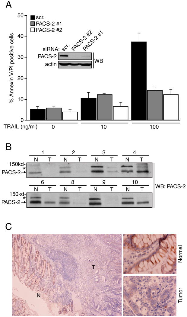
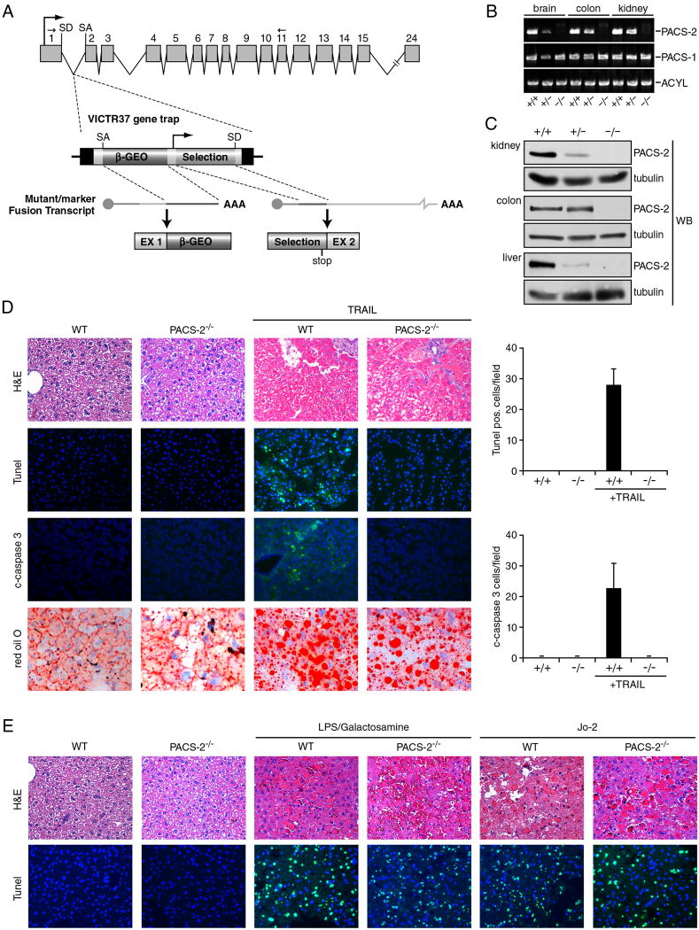
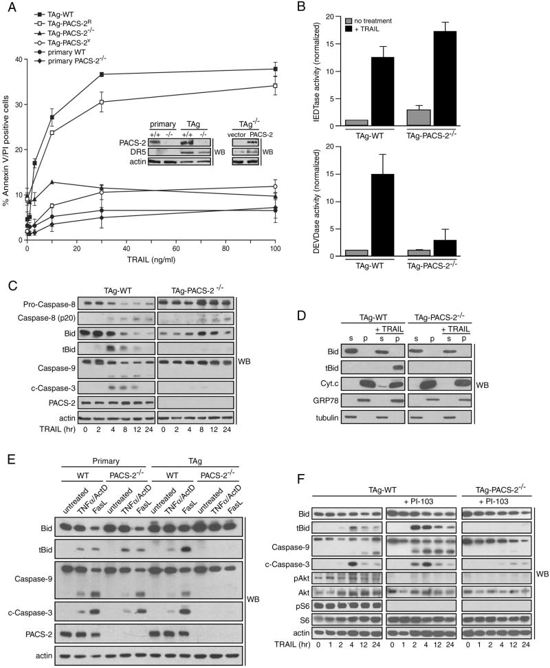
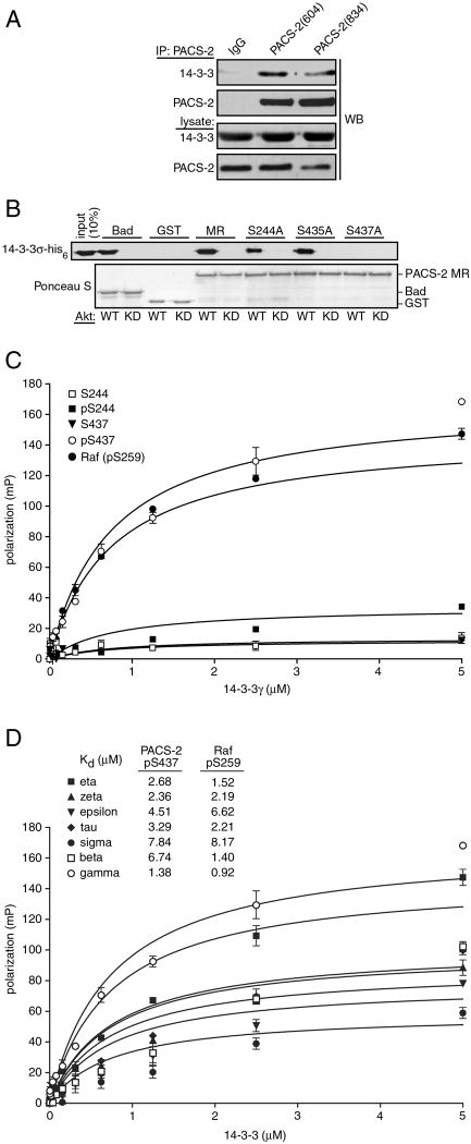
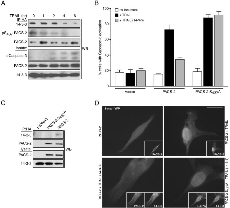
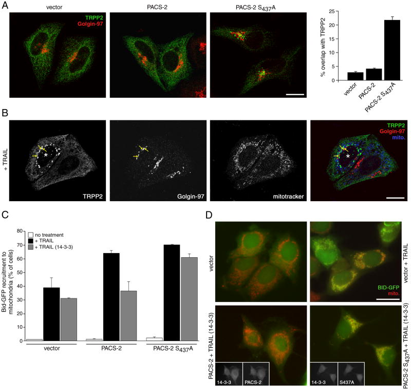
TRAIL selectively kills diseased cells in vivo, spurring interest in this death ligand as a potential therapeutic. However, many cancer cells are resistant to TRAIL, suggesting the mechanism mediating TRAIL-induced apoptosis is complex. Here we identify PACS-2 as an essential TRAIL effector, required for killing tumor cells in vitro and virally infected hepatocytes in vivo. PACS-2 is phosphorylated at Ser437 in vivo, and pharmacologic and genetic studies demonstrate Akt is an in vivo Ser437 kinase. Akt cooperates with 14-3-3 to regulate the homeostatic and apoptotic properties of PACS-2 that mediate TRAIL action. Phosphorylated Ser437 binds 14-3-3 with high affinity, which represses PACS-2 apoptotic activity and is required for PACS-2 to mediate trafficking of membrane cargo. TRAIL triggers dephosphorylation of Ser437, reprogramming PACS-2 to promote apoptosis. Together, these studies identify the phosphorylation state of PACS-2 Ser437 as a molecular switch that integrates cellular homeostasis with TRAIL-induced apoptosis.


Figures

Figure 1
Figure 1. PACS-2 is required for TRAIL-mediated tumor cell death
(A) HCT116 cells were co-nucleofected with pmaxGFP and either a control siRNA (scr.) or PACS-2 siRNAs (#1, Qiagen, #2, Dharmacon) and then treated with 0, 10 or 100 ng/ml TRAIL for 16 hr. GFP+ cells were analyzed for apoptosis by flow cytometry. Data are represented as mean +/- SEM. Insets: Western blot of PACS-2 in control and PACS-2 knockdown cells. (B) Matched colorectal tumor (T) and adjacent normal (N) colonic tissue from eight volunteers with metastatic (samples 1-3) or stage 3 (samples 4,6 8-10) cancer were homogenized and 100 μg of total protein from each sample was analyzed by western blot. Arrow, PACS-2; *, 150 kDa PACS-2 immunoreactive protein of unknown identity. (C) A paraffin block of colonic tumor was sectioned and analyzed for PACS-2 expression by immunohistochemistry. Left, Normal colonic epithelium (N) and adjacent tumor (T). Right, Magnification of normal and tumor regions.
Figure 2
Figure 2. PACS-2 is required for TRAIL-mediated apoptosis in vivo
(A) VICTR37 retroviral gene trap inserted into intron 1 of the murine PACS-2 gene. The gene trap (PACS-2GT(OST426086)) produces a truncated protein chimera containing only the first 40 amino acids of the 889 amino acid PACS-2. Arrows above exons 1 and 11 depict PCR primers used in panel B. SD, splice donor site; SA, splice acceptor. (B) RNA was isolated from PACS-2 WT (PACS-2+/+), heterozygote (PACS-2+/GT(OST426086), hereafter called PACS-2+/-), or PACS-2 gene trap ((PACS-2 GT(OST426086)/GT(OST426086), hereafter called PACS-2-/-) littermates and analyzed by RT-PCR to detect PACS-1, PACS-2 and ATP citrate lyase (ACYL, positive control). (C) Tissues from WT, +/- or -/- mice were analyzed by western blot for PACS-2. (D) WT and PACS-2-/- mice (n = 4 in each group) were injected with AdGFP and mouse AdTrail. TUNEL and c-caspase-3 positive cells were counted in liver sections from WT and PACS-2-/- mice were visualized (magnification, 200x) and quantified as the number of TUNEL- and cleaved caspase 3-positive cells per 20X field (graphs). Liver steatosis was detected by Oil Red O staining. (E) WT and PACS-2-/- mice (n = 4 in each group) were injected with the mAb Jo2 to stimulate Fas-mediated apoptosis or with LPS /GalN to stimulate TNFα-mediated apoptosis and TUNEL-positive nuclei were counted (original magnification 200x).
Figure 3
Figure 3. PACS-2 is required for TRAIL-induced apoptosis of SV40 large T antigen-transformed MEFs
(A) Primary WT and PACS-2-/- MEFs, as well as TAg-WT, TAg-PACS-2-/-, TAg-PACS-2V or TAg-PACS-2R MEFs were treated with increasing concentrations of TRAIL for 16 hr and then analyzed for apoptosis by Annexin V/ propidium iodide staining. Inset, western blot of PACS-2 and DR5 expression in the cultures. Data are represented as mean +/- SEM. (B) Lysates from TAg-WT and TAg-PACS-2-/- MEFs treated or not with 25 ng/ml TRAIL for 4 hr were incubated with the caspase-8 substrate Ac-IETD-AFC or the caspase-3 substrate Ac-DEVD-AFC and free AFC fluorescence was quantified. Data are represented as mean +/- SEM. (C) TAg-WT and TAg-PACS-2-/- MEFs were treated with 50 ng/ml TRAIL for the times indicated and the cleavage of each apoptotic factor was determined by western blot. Actin, loading control. (D) TAg-WT and TAg-PACS-2-/- MEFs were treated or not with 25 ng/ml TRAIL for 4 hr. Cells were permeabilized with digitonin and the cytosol supernatant (S) and membrane pellets (P) were separated by centrifugation. GRP78, organelle pellet marker; tubulin, cytosol marker. (E) Primary or TAg MEFs were treated with 100 ng/ml TNFα plus 1 μg/ml Actinomycin D or 250ng/ml FasL for 4 hr and analyzed as described in panel C. (F) TAg-WT and TAg-PACS-2-/- MEFs were pretreated or not with vehicle (DMSO) or 1 μM PI-103 for 1 hr then treated with 50 ng/ml TRAIL for the indicated times and analyzed as described in panel C.
Figure 4
Figure 4. Akt phosphorylates PACS-2 Ser437 in vitro and vivo
(A) Diagram of PACS-2 showing the N-terminal region (NTR), cargo-binding region (FBR), which contains Ser43, the middle region (MR), which contains Ser244, Ser435 and Ser437, and the C-terminal region (CTR). (B) The indicated GST-fusion proteins were incubated with [γ-32P]-ATP and constitutively active HA-Akt (WT) or kinase-dead HA-Akt (KD). Con, immunoisolation from mock-infected cells. Left, autoradiography; Right, input. (C) The indicated GST-fusion proteins were incubated with [γ-32P]-ATP and immunoisolated HA-Akt as described in panel B. Top, autoradiography; Bottom, input. (D) Peptides from in-gel limited trypsin digestion of HA-tagged PACS-2 were evaluated for candidate phosphopeptide by mass spectrometry. A phosphopeptide at [M+2H]=450.7, corresponding to the PACS-2 tryptic peptide RSTpS437LKER with one phosphorylation, was identified by nanoLC-MS/MS. (E) Left; Brain cytosol from WT and PACS-2-/- mice was incubated with affinity purified anti-PACS-2 (Ab 832) and immunoprecipitated proteins were analyzed by western blot using anti-PACS-2 (Ab 193) or mAb 81 to detect pSer437. Right; Liver and small intestine cytosol from WT mice were analyzed as described above. (F) Lysates from WT and Akt1-/- MEFs were analyzed as described in panel E to detect pSer437. (G) HCT116 cells were harvested in proliferative phase (untreated), serum-starved for 16 hr (starved), starved and refed with 20% serum for 3 hr (control), or starved and pre-treated with 1 μM PI-103, 20 nM RAD011, 1 μM BKM-120 or 20 nM BEZ-235 for one hr prior to refeeding with 20% serum for 3 hr. Cells were analyzed by western blot to detect total PACS-2 and PACS-2 pSer437. The pSer437:total PACS-2 ratio was calculated and normalized to the untreated sample. Data are represented as mean +/- SD.
Figure 5
Figure 5. 14-3-3 proteins bind PACS-2 phospho-Ser437
(A) PACS-2 was immunoprecipitated from rat brain cytosol and co-precipitating 14-3-3 was detected by western blot. (B) The indicated GST fusion proteins were incubated with ATP and constitutively active HA-Akt (WT) or kinase-dead HA-Akt (KD), then incubated with 14-3-3σ-his6. Protein complexes were captured with glutathione-sepharose and bound 14-3-3σ-his6 was detected by western blot. (C) (TMR)–labeled 16mer peptides containing pSer259-Raf, PACS-2 Ser244-PACS-2, pSer244-PACS-2, Ser437-PACS-2 or pSer437-PACS-2 (1 nM) were incubated in triplicate with increasing concentrations of GST-14-3-3γ. Fluorescence polarization signals were recorded and used to calculate Kds. (D) 14-3-3 isoforms were incubated with (TMR)-labeled pSer437-PACS-2 or pSer259-Raf peptides and analyzed as described in panel C. Calculated Kd values are shown.
Figure 6
Figure 6. 14-3-3 regulates the apoptotic activity of PACS-2 Ser437
(A) HCT116 cells expressing HA-tagged PACS-2 and myc-tagged 14-3-3ζ were treated with 25 ng/ml TRAIL for the indicated times. PACS-2ha immunoprecipitates were immunoblotted to detect co-precipitating 14-3-3ζ and pSer437. (B) TAg-PACS-2-/- cells were co-transfected with an eYFP-tagged caspase-3 reporter together with pcDNA3.1 (vector) or plasmids expressing HA-tagged PACS-2 or PACS-2S437A alone or with 14-3-3ζ-myc. Top: Cells treated with 10 ng/ml mouse TRAIL for 16 hr were fixed and the number of nuclear eYFP-positive cells indicating activated caspase-3 were analyzed. Data are represented as mean +/- SEM. Bottom: Representative immunofluorescence images of the quantified data. Insets, co-expression of 14-3-3ζ-myc and HA-tagged PACS-2 or PACS-2S437A. Scale bar, 20 μm. (C) TAg-PACS-2-/- cells described in B were lysed, PACS-2 immunoprecipitated and co-precipitating 14-3-3ζ-myc detected by western blot.
Figure 7
Figure 7. PACS-2 Ser437 and 14-3-3 integrate membrane traffic with apoptotic Bid action
(A) HeLa cells were nucleofected (Amaxa) with myc-TRPP2 together with either empty vector or HA-tagged PACS-2 or PACS-2S437A. After 36 hr, the cells were fixed and processed for confocal microscopy using anti-myc (mAb 9E10, green) and anti-Golgin-97 (red) and visualized using Alexa-conjugated secondary antibodies. Right: Morphometric analysis. Error bars represent mean +/- SEM. Scale bar, 20 μm. (B) HeLa cells expressing myc-TRPP2 were treated with 20 ng/ml TRAIL for 4 hr and processed for confocal microscopy. Mitotracker (pseudocolored blue), anti-myc (green) and anti-Golgin-97 (red) are shown. *, apoptotic cell showing puncate myc-TRPP2 and collapsed mitochondria. Arrows, overlap of myc-TRPP2 and Golgin-97. Scale bar, 20 μm. (C) MCF-7:Bid-GFP cells were nucleofected (Amaxa) with pcDNA3.1 (vector) or plasmids expressing HA-tagged PACS-2 or PACS-2S437A alone or with 14-3-3ζ-myc. The cells were then treated with vehicle or 20 ng/ml TRAIL for 4 hr. Left: The percent of cells containing Bid-GFP translocated to mitochondria (mitotracker, red) in random fields (300 cells minimum) were quantified. Data are represented as mean +/- SD. Right: Immunofluorescence of MCF-7:Bid-GFP expressing the indicated proteins and treated or not with TRAIL. Insets, co-expression of 14-3-3ζ-myc with HA-tagged PACS-2 or PACS-2S437A. Scale bar, 20 μm.

References

    1. Anderson GR, Brenner BM, Swede H, Chen N, Henry WM, Conroy JM, Karpenko MJ, Issa JP, Bartos JD, Brunelle JK, et al. Intrachromosomal genomic instability in human sporadic colorectal cancer measured by genome-wide allelotyping and inter-(simple sequence repeat) PCR. Cancer Res. 2001;61:8274–8283. - PubMed
    1. Ashkenazi A, Herbst RS. To kill a tumor cell: the potential of proapoptotic receptor agonists. J Clin Invest. 2008;118:1979–1990. - PMC - PubMed
    1. Aslan JE, Thomas G. Death by committee: Organellar Trafficking and Communication in Apoptosis. Traffic. 2009 in press. - PMC - PubMed
    1. Atkins KM, Thomas L, Youker RT, Harriff MJ, Pissani F, You H, Thomas G. HIV-1 Nef Binds PACS-2 to Assemble a Multikinase Cascade That Triggers Major Histocompatibility Complex Class I (MHC-I) Down-regulation: ANALYSIS USING SHORT INTERFERING RNA AND KNOCK-OUT MICE. J Biol Chem. 2008;283:11772–11784. - PMC - PubMed
    1. Chen X, Thakkar H, Tyan F, Gim S, Robinson H, Lee C, Pandey SK, Nwokorie C, Onwudiwe N, Srivastava RK. Constitutively active Akt is an important regulator of TRAIL sensitivity in prostate cancer. Oncogene. 2001;20:6073–6083. - PubMed

Publication types

MeSH terms