. 2009 Jul 24:1281:1-14.
doi: 10.1016/j.brainres.2009.05.050. Epub 2009 May 28.

Extracellular norepinephrine, norepinephrine receptor and transporter protein and mRNA levels are differentially altered in the developing rat brain due to dietary iron deficiency and manganese exposure

Affiliations

Extracellular norepinephrine, norepinephrine receptor and transporter protein and mRNA levels are differentially altered in the developing rat brain due to dietary iron deficiency and manganese exposure

Joel G Anderson et al. Brain Res. .

Abstract

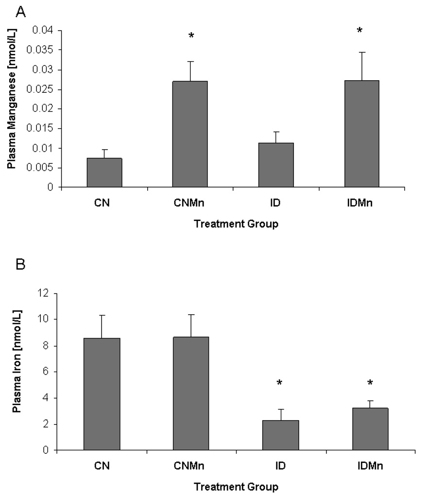
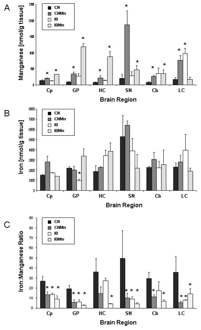
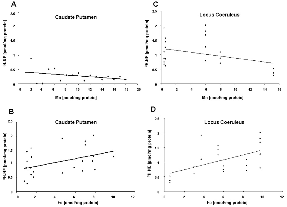
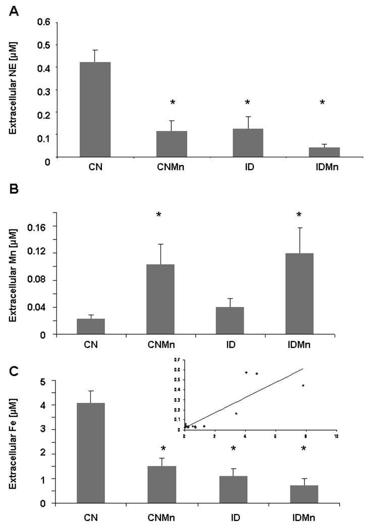
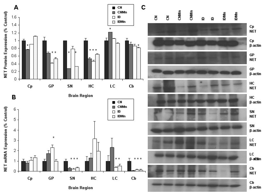
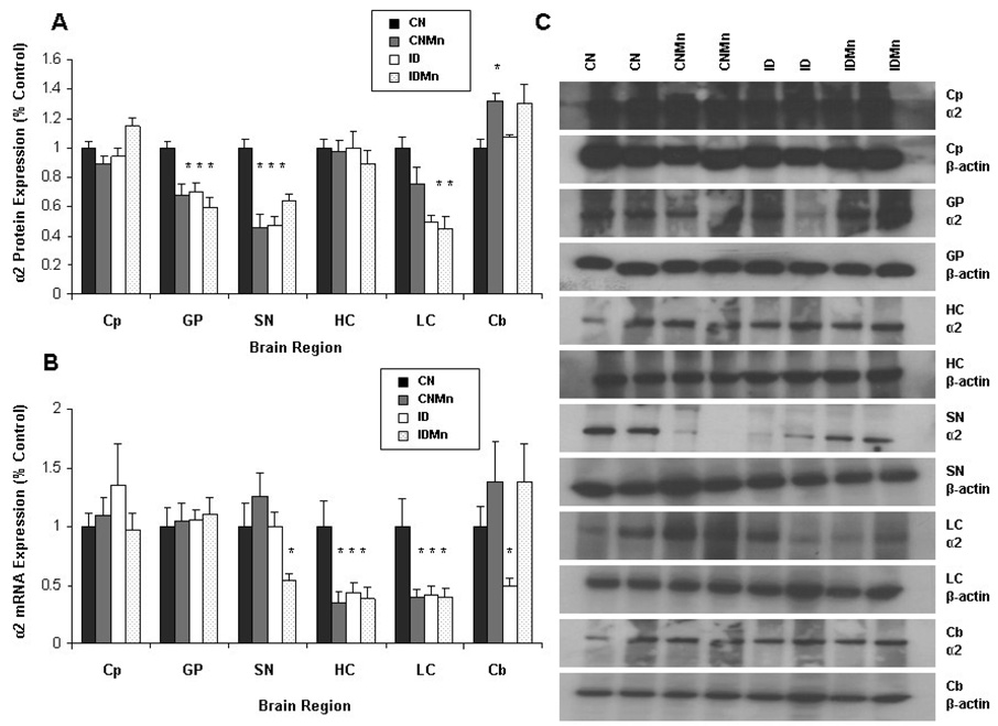
Manganese (Mn) is an essential trace element, but overexposure is characterized by Parkinson's like symptoms in extreme cases. Previous studies have shown that Mn accumulation is exacerbated by dietary iron deficiency (ID) and disturbances in norepinephrine (NE) have been reported. Because behaviors associated with Mn neurotoxicity are complex, the goal of this study was to examine the effects of Mn exposure and ID-associated Mn accumulation on NE uptake in synaptosomes, extracellular NE concentrations, and expression of NE transport and receptor proteins. Sprague-Dawley rats were assigned to four dietary groups: control (CN; 35 mg Fe/kg diet), iron-deficient (ID; 6 mg Fe/kg diet), CN with Mn exposure (via the drinking water; 1 g Mn/L) (CNMn), and ID with Mn (IDMn). (3)H-NE uptake decreased significantly (R=-0.753, p=0.001) with increased Mn concentration in the locus coeruleus, while decreased Fe was associated with decreased uptake of (3)H-NE in the caudate putamen (R=0.436, p=0.033) and locus coeruleus (R=0.86; p<0.001). Extracellular concentrations of NE in the caudate putamen were significantly decreased in response to Mn exposure and ID (p<0.001). A diverse response of Mn exposure and ID was observed on mRNA and protein expression of NE transporter (NET) and alpha(2) adrenergic receptor. For example, elevated brain Mn and decreased Fe caused an approximate 50% decrease in NET and alpha(2) adrenergic receptor protein expression in several brain regions, with reductions in mRNA expression also observed. These data suggest that Mn exposure results in a decrease in NE uptake and extracellular NE concentrations via altered expression of transport and receptor proteins.


Figures

Figure 1
Figure 1
Plasma metal concentrations at six weeks. (A) Plasma manganese concentrations expressed as nmol/L were significantly increased in those animals receiving manganese supplementation versus those animals receiving deionized water alone (p=0.02) (n=24). (B) A significant decrease in plasma iron concentration was observed in animals receiving the ID diet versus the CN diet (p=0.007).
Figure 2
Figure 2
Brain metal concentrations at six weeks. Overall, Mn exposure caused a significant increase in brain regional Mn concentrations versus CN. Mean concentrations ± SEM are shown for manganese (A) and iron (B) for caudate putamen (Cp), globus pallidus (GP), hippocampus (HC), substantia nigra (SN), cerebellum (Cb), and locus coeruleus (LC). The Fe:Mn ratio is also reported, illustrating the impact of Mn exposure on Fe homeostasis (C). CN is represented in black (n=6), CNMn in gray (n=6), ID in white (n=6), and IDMn in dotted area (n=6). Asterisks (*) indicate a statistically significant difference from CN according to Dunnett’s post-hoc analysis.
Figure 3
Figure 3
Effect of dietary treatment on uptake of 3H-NE. 3H-NE uptake decreased with increased Mn concentration in the locus coeruleus, with decreased Fe associated with decreased uptake of 3H-NE in the caudate putamen and locus coeruleus. Correlational analysis of 3H-NE uptake in the caudate putamen versus (A) synaptosomal Mn concentration and (B) synaptosomal Fe concentration (R=0.436; p=0.033). Correlation analysis of 3H-NE uptake in the locus coeruleus versus (C) synaptosomal Mn concentration (R=−0.753; p=0.001) and (D) synaptosomal Fe concentration (R=0.86; p<0.001) (n=24).
Figure 4
Figure 4
Microdialysate analysis. Extracellular concentrations of NE in the caudate putamen were significantly decreased in response to Mn exposure and ID. Mean concentrations ± SEM are shown for (A) extracellular NE, (B) manganese, and (C) iron in microdialysate samples from the caudate putamen after six weeks of dietary treatment (n=24). Inset: Correlational analysis of extracellular NE versus extracellular Fe (R=0.86; p<0.001). *p<0.001
Figure 5
Figure 5
Effect of dietary treatment on NET protein and mRNA expression. Overall, Mn exposure and ID lead to a decrease in NET protein and mRNA expression. Mean expression as percentage of control ± SEM for NET (A) protein and (B) mRNA relative to β-actin are shown for caudate putamen (Cp), globus pallidus (GP), hippocampus (HC), substantia nigra (SN), locus coeruleus (LC), and cerebellum (Cb) (n=24). CN is represented in black, CNMn in gray, ID in white, and IDMn in dotted area. (C) Representative blots for NET and β-actin for each region are shown, with each band representing an individual animal. *p<0.05 according to Dunnett’s post-hoc analysis
Figure 6
Figure 6
Effect of dietary treatment on α2 receptor protein and mRNA expression. Overall, Mn exposure and ID lead to a decrease in α2 receptor protein and mRNA expression. Mean expression as percentage of control ± SEM for α2 receptor (A) protein and (B) mRNA relative to β-actin are shown for caudate putamen (Cp), globus pallidus (GP), hippocampus (HC), substantia nigra (SN), locus coeruleus (LC) and cerebellum (Cb) (n=24). CN is represented in black, CNMn in gray, ID in white, and IDMn in dotted area. (C) Representative blots for α2 receptor and β-actin for each region are shown, with each band representing an individual animal. *p<0.05 according to Dunnett’s post-hoc analysis
Figure 7
Figure 7
NE biology during Mn overload and reduced Fe. This simple schematic of the basal ganglia represents the potential cause and consequences of the decreased extracellular NE concentrations in the striatum (caudate putamen) due to alterations of Mn and Fe status observed in the current study. (1) Altered expression of NE transport and receptor proteins and/or neuronal loss in the locus coeruleus (LC) would lead to a decrease in NE release (thin dotted line), (2) decreasing extracellular NE concentrations in the striatum, reducing the activity of the GABA striatopallidal projection neurons (3) (dotted line). This reduction in activity would (4) increase the GABAergic inhibitory firing from the globus pallidus (GP) to the subthalamic nucleus (STN) (heavy black line), in turn (5) decreasing the excitatory glutamatergic firing from this region to the substantia nigra (SN) (dotted line). (6) Decreased glutamatergic excitation in the SN, along with decreased GABAergic inhibition from the striatonigral projection neurons (dotted line), decreased adrenergic activity from the LC (1) (thin dotted line), and decreased protein expression of NET and α2 adrenergic receptor, would lead to a dysregulation of dopaminergic firing to the Cp along the nigrostriatal pathway (alternating line).

References

    1. Anderson JG, Fordahl SC, Cooney PT, Erikson KM. Manganese exposure alters extracellular GABA, GABA receptor and transporter protein and mRNA levels in the developing rat brain. NeuroToxicology. 2008;29:1044–1053. - PMC - PubMed
    1. Anderson JG, Cooney PT, Erikson KM. Brain manganese accumulation is inversely related to GABA uptake in male and female rats. Tox. Sci. 2007;95:188–195. - PubMed
    1. Andrade R, Aghajanian GK. Locus coeruleus activity in vitro: intrinsic regulation by a calcium-dependent potassium conductance but not α2-adrenoceptors. J Neurosci. 1984;4:161–170. - PMC - PubMed
    1. Aschner M, Erikson KM, Dorman DC. Manganese dosimetry: species differences and implications for neurotoxicity. Crit. Rev. Tox. 2005;35:1–32. - PubMed
    1. Autissier N, Rochette L, Dumas P, Beley A, Loireau A, Bralet J. Dopamine and norepinephrine turnover in various regions of the rat brain after chronic manganese chloride administration. Toxicology. 1982;24:175–182. - PubMed

Publication types

MeSH terms