. 2009 Sep;30(25):4085-93.
doi: 10.1016/j.biomaterials.2009.04.051. Epub 2009 May 29.

Promoting angiogenesis via manipulation of VEGF responsiveness with notch signaling

Affiliations

Promoting angiogenesis via manipulation of VEGF responsiveness with notch signaling

Lan Cao et al. Biomaterials. 2009 Sep.

Abstract

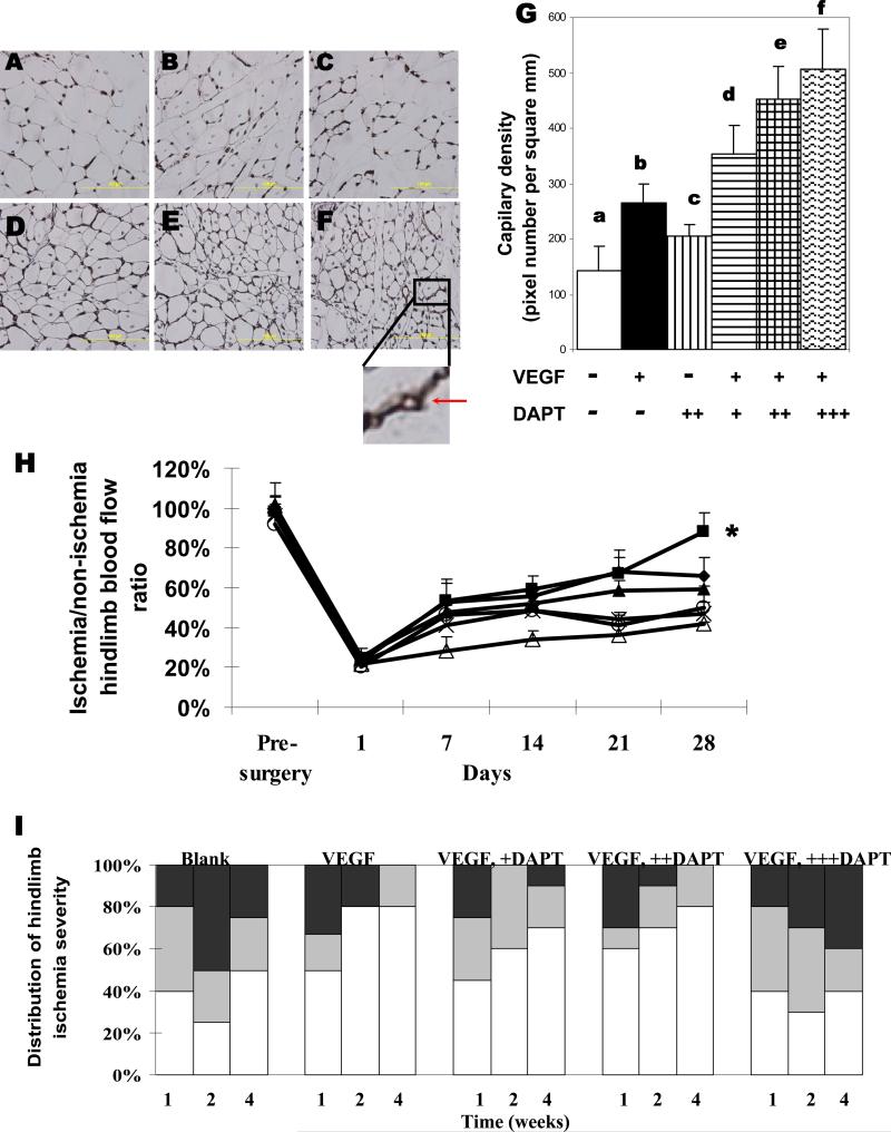
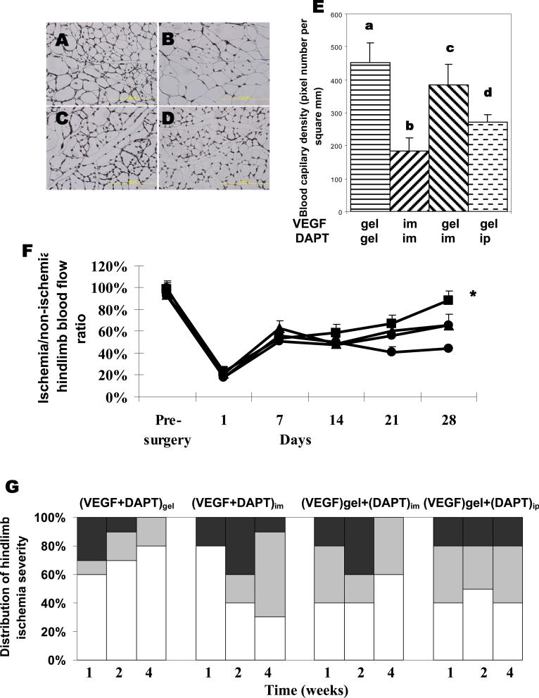
Promoting angiogenesis via delivery of vascular endothelial growth factor (VEGF) and other angiogenic factors is both a potential therapy for cardiovascular diseases and a critical aspect for tissue regeneration. The recent demonstration that VEGF signaling is modulated by the Notch signaling pathway, however, suggests that inhibiting Notch signaling may enhance regional neovascularization, by altering the responsiveness of local endothelial cells to angiogenic stimuli. We tested this possibility with in vitro assays using human endothelial cells, as well as in a rodent hindlimb ischemia model. Treatment of cultured human endothelial cells with DAPT, a gamma secretase inhibitor, increased cell migration and sprout formation in response to VEGF stimulation with a biphasic dependence on DAPT concentration. Further, delivery of an appropriate combination of DAPT and VEGF from an injectable alginate hydrogel system into ischemic hindlimbs led to a faster recovery of blood flow than VEGF or DAPT alone; perfusion levels reached 80% of the normal level by week 4 with combined DAPT and VEGF delivery. Direct intramuscular or intraperitoneal injection of DAPT did not result in the same level of improvement, suggesting that appropriate presentation of DAPT (gel delivery) is important for its activity. DAPT delivery from the hydrogels also did not lead to any adverse side effects, in contrast to systemic introduction of DAPT. Altogether, these results suggest a new approach to promote angiogenesis by controlling Notch signaling, and may provide new options to treat patients with diseases that diminish angiogenic responsiveness.


Figures

Figure 1
Figure 1
Effect of DAPT on endothelial cell phenotype in vitro. (A) Fractional change in cell number for cells cultured in the media supplemented with (+) or without (−) 10ng/mL VEGF, and without (−) or with 0.25μM(+), 2.5 μM (++), or 25μM (+++) DAPT. (B) Fractional change in cell number for cells exposed to surface-coated 1μg/mL Dll4 (− − + +), Dll4 with 10ng/mL VEGF (− + + +), or Dll4, VEGF and 0.25μMDAPT (− − − +). Cell number ratio was calculated based on the number of seeded cells. (C) Cell migration towards medium containing VEGF and HGF (each 50ng/mL) as influenced by 0.25μM (+), 2.5μM (++) or 25μM (+++) DAPT. (D) Sprout formation of endothelial cells, normalized to the total bead number. Mean values are presented with standard deviations (n=4–6). * represents P<0.05 as compared to other noted conditions. ** represents P<0.05 as compared to all other conditions. NS, not statistically significant.
Figure 2
Figure 2
Effects of VEGF and DAPT exposure on VEGFR2 expression of endothelial cells. Cells were untreated (control), or treated with 10ng/mL VEGF, 2.5μM DAPT, or a combination of both (VEGF+DAPT) for 15min. Cell lysates extracted using either lysis buffer A (LyA), or lysis buffer B (LyB) buffer that extract all cellular components, were examined for the different treatment conditions and analyzed using western blotting. A control protein, vinculin was also shown for comparison.
Figure 3
Figure 3
In vitro factor release. DAPT release was analyzed from alginate hydrogels containing 86ng DAPT alone (diamond), 860ng DAPT alone (square), 8600ng DAPT alone (triangle), or 860ng DAPT and 3μg VEGF (horizontal line). VEGF release was monitored from gels containing 3μg VEGF alone (star). Mean values are presented with standard deviations (n=6).
Figure 4
Figure 4
Analysis of angiogenesis in ischemic hindlimbs. Representative images (A-F) and quantities of capillaries in muscle tissues adjacent to the injection site, measured from tissues immunostained for CD31 (G). Tissues were retrieved from animals treated with either blank gels (A, a), VEGF (3μg) (B, b), 860ng DAPT(C, c), VEGF+86ng DAPT (D, d), VEGF+860ng DAPT (E, e), or VEGF+8600ng DAPT (F, f) delivered from alginate hydrogels. The insert shows the lumen structure of a positively stained capillary. Scale bar, 100μm. (H) Blood perfusion profile of hindlimbs treated with alginate hydrogels incorporating 860ng DAPT and 3μg VEGF (square), 86ng DAPT+VEGF (diamond),, VEGF alone (triangle), 860ng DAT alone (open circle), 8600ng DAPT +VEGF (star), or blank gels (open triangle). Mean values are presented with standard deviations (n=6). *, statistically significant difference (P<0.05), as compared to other conditions. (I) Quantification and distribution of hindlimb ischemia severity observed in different experimental groups over time. Tissue necrosis of hindlimbs subjected to surgery were visually examined, and grouped as normal (displaying limb integrity from non-ischemic hindlimbs of the same animal, white regions), or presenting one necrotic toe (gray), or multiple necrotic toes (black). Animals were treated with blank gels, 3μg VEGF, or combinations of VEGF with 86ng DAPT (+DAPT), 860ng DAPT (++DAPT), or 8600ng DAPT (+++DAPT).
Figure 5
Figure 5
Effects of the delivery method of DAPT on angiogenesis in vivo. Representative images (A-D) and quantities of capillaries in tissues adjacent to the injection site measured by CD31 immunostaining of tissue samples (E). Tissues were retrieved from animals treated with gel delivery of both VEGF and DAPT(A, a), intramuscular injection of VEGF and DAPT (B, b), or gel delivery of VEGF combined with intramuscular injection of DAPT (C, c), or combined with intraperitoneal injection of DAPT (D, d). The doses are 860ng for DAPT and 3μg for VEGF respectively. Scale bar, 100μm. (F) Blood perfusion profile of hindlimbs treated with gel delivery of VEGF and DAPT (diamond), gel delivery of VEGF and intramuscular delivery of DAPT (triangle), gel delivery of VEGF and intraperitoneal delivery of DAPT (square), or intramuscular delivery of DAPT and VEGF (circle). Mean values are presented with standard deviations (n=6). *, statistically significant difference (P<0.05), as compared to other conditions. (G) Quantification and distribution of hindlimb ischemia severity observed in different experimental groups over time. Tissue necrosis of hindlimbs subjected to surgery were visually examined, and grouped as normal (displaying limb integrity from non-ischemic hindlimbs of the same animal, white regions), or presenting one necrotic toe (gray), or multiple necrotic toes (black). Animals were treated with gel delivery of VEGF and DAPT ((VEGF+DAPT)gel), gel delivery of VEGF and intramuscular delivery of DAPT ((VEGF)gel+(DAPT)im), gel delivery of VEGF and intraperitoneal delivery of DAPT ((VEGF)gel+(DAPT)ip), or intramuscular delivery of DAPT and VEGF ((VEGF+DAPT)im).
Figure 6
Figure 6
Effect of DAPT delivery method on the structure and phenotype of small intestine. Representative images of tissue sections from small intestines isolated from mice without any treatment(control), mice treated with 860ng DAPT delivered from alginate gel ((DAPT)gel), or 860ng DAPT injected intraperitoneally ((DAPT)ip), which were stained against Notch gene HES-1 expression (A, B, C), Ki67 expression (D, E, F), alcian blue staining for glycosaminoglycans (G, H, I), or hematoxylin and eosin staining (J, K, L). Scale bar, 100μm.

References

    1. Carmeliet P. Angiogenesis in life, disease and medicine. Nature. 2005;438:932–936. - PubMed
    1. Cao L, Mooney DJ. Spatiotemporal control over growth factor signaling for therapeutic neovascularization. Adv Drug Deliv Rev. 2007;59(13):1340–1350. - PMC - PubMed
    1. Laschke MW, Harder Y, Amon M, Martin I, Farhadi J, Ring A, et al. Angiogenesis in tissue engineering: breathing life into constructed tissue substitutes. Tissue Eng. 2006;12(8):2093–2104. - PubMed
    1. Brey EM, Uriel S, Greisler HP, McIntire LV. Therapeutic neovascularization: contributions from bioengineering. Tissue Eng. 2005;11(3–4):567–584. - PubMed
    1. Ferrara N. Vascular endothelial growth factor: basic science and clinical progress. Endocr Rev. 2004;25(4):581–611. - PubMed

Publication types

MeSH terms