. 2009 Jul 31;390(5):991-1006.
doi: 10.1016/j.jmb.2009.05.072. Epub 2009 May 29.

RNA chaperones stimulate formation and yield of the U3 snoRNA-Pre-rRNA duplexes needed for eukaryotic ribosome biogenesis

Affiliations

RNA chaperones stimulate formation and yield of the U3 snoRNA-Pre-rRNA duplexes needed for eukaryotic ribosome biogenesis

Tímea Gérczei et al. J Mol Biol. .

Abstract

Short duplexes between the U3 small nucleolar RNA and the precursor ribosomal RNA must form quickly and with high yield to satisfy the high demand for ribosome synthesis in rapidly growing eukaryotic cells. These interactions, designated the U3-ETS (external transcribed spacer) and U3-18S duplexes, are essential to initiate the processing of small subunit ribosomal RNA. Previously, we showed that duplexes corresponding to those in Saccharomyces cerevisiae are only observed in vitro after addition of one of two proteins: Imp3p or Imp4p. Here, we used fluorescence-based and other in vitro assays to determine whether these proteins possess RNA chaperone activities and to assess whether these activities are sufficient to satisfy the duplex yield and rate requirements expected in vivo. Assembly of both proteins with the U3 small nucleolar RNA into a chaperone complex destabilizes a U3 stem structure, apparently to expose its 18S base-pairing site. As a result, the chaperone complex accelerates formation of the U3-18S duplex from an undetectable rate to one comparable with the intrinsic rate observed for hybridizing short duplexes. The chaperone complex also stabilizes the U3-ETS duplex by 2.7 kcal/mol. These chaperone activities provide high U3-ETS duplex yield and rapid U3-18S duplex formation over a broad concentration range to help ensure that the U3-precursor ribosomal RNA interactions limit neither ribosome biogenesis nor rapid cell growth. The thermodynamic and kinetic framework used is general and thus suitable for investigating the mechanism of action of other RNA chaperones.


Figures

Fig. 1
Fig. 1
A schematic view of U3-pre-rRNA interactions from S. cerevisiae. (a) The U3 snoRNA (thick black line) base pairs with the pre-rRNA that embeds three mature rRNAs (grey) between internal and external transcribed spacers (thin line). Formation of the U3-ETS and U3-18S duplexes is a prerequisite for the U3-dependent cleavage events at A0, A1 and A2. (b) Framework for formation of the U3-ETS and U3-18S duplexes with the minimal U3 binding site for Imp3p and Imp4p, U3 MINI. Duplex yield is limited by the thermodynamic instability of the U3-ETS duplex. Formation of the U3-18S duplex is hindered by a kinetic unfolding barrier; the box A/A’ stem structure of U3 MINI must open up to expose the base pairing site. Thus, formation of the U3-18S duplex involves two steps: unfolding U3 MINI to U3 MINI* and hybridization. The duplex dissociation constant (Kd), duplex association and dissociation rate constants (kon and koff) and the equilibrium constant between U3 MINI and U3 MINI*, Keq, are shown. The (ETS) or (18S) suffix is added to distinguish U3-ETS duplex parameters from those of the U3-18S duplex. Coloring of ETS (cyan) and 18S (red) sequences is used henceforth.
Fig. 2
Fig. 2
Assembly of the chaperone complex between Imp3p, Imp4p and U3 MINI is RNA-dependent and may be ordered. (a) EFRET values are shown for three conditions: addition of Fl-Imp3p to a preincubated mixture of Rh-Imp4p and U3 MINI (filled bar); a mixture of Fl-Imp3p and Rh-Imp4p (hashed bar); and addition of Rh-Imp4p to a preincubated mixture of Fl-Imp3p and U3 MINI (open bar). (b) An emission spectrum of Rh-Imp4p added to a preincubated mixture of Fl-Imp3p and U3 MINI (solid line) illustrates the decrease in Fl emission (down arrow) with concomitant increase in Rh emission (up arrow) of a FRET signal. The emission spectrum of a preformed U3/Fl-Imp3p binary complex was the same in the presence (dashed line) and absence (data not shown) of unlabeled Imp4p, verifying that the signal is a result of FRET and not fluorescence quenching by Imp4p binding. (c) A schematic overview of the three steps of purification of the chaperone complex via metal affinity resin: (i) His6-Imp3p (grey), Imp4p (white), U3 snoRNA (black) or some combination thereof are loaded (L) onto a metal affinity resin; (ii) the column is washed; and (iii) the eluant is eluted (E) with addition of imidazole. (d) Four L and E fractions were analyzed on SDS-PAGE stained with ethidium bromide and silver nitrate to visualize the U3 snoRNA and the proteins, respectively.
Fig. 3
Fig. 3
Free energy reaction profiles illustrate the six possible mechanisms used by proteins to mediate duplex formation by changing the energy levels of the substrate, transition state, product or some combination thereof. Evaluation of how the magnitude of the duplex kon, koff and Kd changes after addition of protein is used to discriminate between alternate mechanisms.
Fig. 4
Fig. 4
The stable box A/A’ stem structure of U3 MINI is unfolded to U3 MINI* by addition of protein, primarily Imp3p. (a) OD260 values for unlabeled U3 MINI melted in the forward (circles) and refolded in the reverse direction (squares). A smoothed derivative plot (dashed line) of the forward melt indicates a Tm of 54 °C, with an enthalpy of 39 kcal/mol and a ΔG°20 °C of 4 kcal/mol. (b) To monitor distance changes at the base of the box A/A’ stem structure trFRET was performed using a doubly labeled substrate: Fl is attached to the 5’ end of U3 MINI via a six carbon linker and an internal Rh label is attached via a longer succinimide linker to C5 of uracil 38. (c) FRET distance distributions between the donor and acceptor of Fl-U3 MINI-Rh upon binding of Imp3p, Imp4p, and 18S as determined by trFRET (Supplemental Fig. S3 contains decay curves). In the absence of protein ~93% fraction of molecules show a distance distribution centered around 19 Å (grey line), with a full width at half-maximum (fwhm) of 18 Å. The fwhm reflects in part the intrinsic flexibility of the RNA in solution. A smaller ~7% fraction (dashed grey line) has distance distribution centered around 45 Å with an fwhm of 37 Å and is likely to result from a small population of U3 MINI dimer. Using an electrophoretic mobility shift assay, the inset shows that 4 ± 2 % of U3 MINI exists in a dimer at the 0.5 µM concentration used for the trFRET studies. Upon binding of Imp3p (dashed black line), a single distance distribution is obtained, centered at a distance of ~32 Å with a fwhm of 8 Å. Subsequent binding of Imp4p (dotted black line) and 18S (solid black line) shows no significant additional change in distance distribution (mean distance of 33 Å with an fwhm of 9 Å and mean distance of 34 Å with an fwhm of 10 Å, respectively). (d) The simplest model indicates that the unfolded RNA nucleotides loop back to permit the separation distance to be independent of Imp4p and 18S binding. Schematics illustrate chaperone complex unfolding the box A/A’ stem of U3 MINI from an A-form helix with a FRET pair separated by 19 Å (left panel) to U3 MINI* where the separation increases to ~32 Å upon binding of Im3p3 and Imp4p (middle panel). Addition of 18S results in a negligible increase in the separation of the FRET pair (right panel).
Fig. 5
Fig. 5
Hybridization kinetics of the U3-18S duplex reveal that kon (18S) does not limit duplex formation. (a) The substrates used for kinetic measurements were the 5’-Fl labeled U3 MINI (Fl-U3 MINI) and the 3’-Rh labeled 18S (18S-Rh). The dashed box reflects MINI-17-18S duplex (Supplemental Fig. S4). (b) Representative normalized fluorescence data are shown for ssFRET dependent fluorescein quenching (10 nM Fl-U3 MINI) in the absence and presence of proteins upon addition of 18S-Rh (10 nM). In the absence of protein (grey circles) the trace is indistinguishable from that observed upon addition of free Rh (10 nM) (open circles) to Fl-U3 MINI, which corresponds to photobleaching. Indistinguishable traces were observed for 1 µM concentrations of substrate as well as when unlabeled 18S was added to Fl-U3 MINI in the presence or absence of protein (data not shown). In contrast, a marked quenching is observed in the presence of saturating amounts of proteins (black squares). (c) Raw representative trace (left y-axis, grey points) is shown for fluorescein quenching upon addition of 18S-Rh to the Fl-U3 MINI/Imp3p/Imp4p complex to achieve a final concentration of 5 nM. The [AB]apparent values calculated using equation (1) are plotted on the right y-axis (black squares) along with the fit (white line) to equation (2a) to give a kon (18S) of 7 × 10⁵ M−1 s−1 (the fit was performed using molar [AB]apparent). (d) Pseudo first-order rates measured with stopped-flow device are plotted for increasing concentrations of 18S-Rh (≥ 150 nM) mixed with 38 nM Fl-U3 MINI in the presence of saturating amounts of protein. The inset shows a representative trace (150 nM 18S-Rh) of fluorescein quenching with kobs calculated by fitting decay traces to equation (4). The kon (18S) value determined in (c) is within the 95% confidence interval of the linear regression fit of the kobs vs 18S-Rh data, which gave a kon (18S) of 5 × 10⁵ M−1 s−1.
Fig. 6
Fig. 6
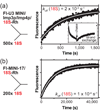
Assembly of the chaperone complex increases koff (18S). (a) Representative trace is shown for a chase initiated with 500-fold excess (5 µM) of unlabeled 18S to a preformed duplex between 10 nM Fl-U3 MINI and 10 nM 18S-Rh in the presence of saturating amounts of Imp3p and Imp4p. Inset shows the decrease in fluorescence due to ssFRET upon hybridization with 18S-Rh, followed by recovery of fluorescence, upon addition of unlabeled 18S, as chase is initiated. Fitting the time dependent increase in fluorescence to equation (5) gave a koff (18S) of 2 × 10⁻³ s−1. (b) Representative chase trace is initiated by addition of unlabeled 18S (0.5 µM) to a preformed complex between Fl-MINI-17 (5 nM) and 18S-Rh (5 nM) in the absence of protein (koff (18S) = 1 × 10⁻⁴ s−1).
Fig. 7
Fig. 7
Reaction profiles illustrating a model for how protein binding accelerates U3-18S duplex formation by unfolding U3 MINI to U3 MINI* (the first step) rather than stimulating hybridization (the second step). In the absence of protein (dashed grey line), the 4 kcal/mol needed to unfold U3 MINI (Fig. 4a) limits the quantities of U3 MINI* and thus reduces the subsequent product duplex to an undetectable level. In contrast, protein binding (black line) unfolds U3 MINI to form a stable U3 MINI* (Fig. 4c), thereby allowing the hybridization step to occur at the intrinsic rate constant for hybridizing short duplexes (~106 M−1s−1) with energetically favorable transitions from U3 MINI to U3 MINI* to duplex. Because hybridization of the MINI-17-18S duplex in the absence of protein (green dotted line) and of the U3-18S duplex in the presence of protein (black line) share a common rate-limiting step, comparison of these reactions is useful. Superimposing the energy of MINI-17 (asterisk) and of the chaperone complex shows the same barrier for the forward reaction (identical kon (18S) values); however, a 20-fold increase in koff (18S) by the chaperone complex, results in 1.7 kcal/mol of product destabilization (Table 1). The energy levels thus differ for the protein bound U3-18S duplex and the protein free MINI-17-18S duplex.
Fig. 8
Fig. 8
Chaperone complex stabilizes the U3-ETS duplex without affecting the association rate constant (kon (ETS)). Representative binding data from electrophoretic mobility shift assays for 32P-ETS-U3 MINI duplex in the absence (a) and presence of the chaperone complex (b). Fraction bound is calculated based on the fraction of 32P-ETS shifted and plotted against increasing concentration of either U3 MINI or the chaperone complex. Kd (ETS) is calculated by fitting fraction bound to equation (6). (c) The fluorescently labeled U3-ETS duplex substrate used for kinetic assays includes the donor 3’-Fl labeled U3 MINI (U3 MINI-Fl) and the acceptor 5’-Rh labeled ETS (Rh-ETS). (d) Representative trace for measurement of kon (ETS) for 5 nM U3 MINI-Fl and 5 nM Rh-ETS in the absence of protein. The fractional change in ssFRET dependent donor quenching is proportional to [AB]apparent in equation (1); kon (ETS) is calculated using equation (2a) (the fit was performed using molar [AB]apparent).
Fig. 9
Fig. 9
Reaction profile illustrating a product stabilization model for the U3-ETS duplex mediated by Imp3p and Imp4p in the chaperone complex. The reaction proceeds from U3 MINI (on the left) to the U3-ETS duplex (on the right) with the same barrier height because kon values remain unchanged in the absence (dashed line) and presence of Imp3p and Imp4p (continuous line). In the proposed product stabilization mechanism formation of the U3-ETS duplex occurs spontaneously. Once formed, the duplex binds tightly into a pocket created by Imp3p and Imp4p to increase duplex stability. The free energy difference between the reactants (free RNAs) and that of the U3-ETS duplexes in the presence and absence of the chaperone complex assumes a standard state of 100 nM and 20 °C and uses Kd (ETS) values from Table 2.
Fig. 10
Fig. 10
The chaperone complex ensures rapid hybridization and a high duplex yield for the U3-pre-rRNA duplexes. (a) U3-18S hybridization is a pre-requisite for cleavage and release of pre-r18S, which has a half-life of 85 s in vivo. In the presence of proteins, the half-life for hybridization is faster than 85 s, for all concentrations greater than 7 nM thus satisfying cellular requirements, calculated using equation (3) and values from Table 1. (b) The percent yield of the U3-ETS duplex (using equation (7) and values from Table 2) in the absence of protein is below 90 % for substrate concentrations less than 63 µM. (c) In the presence of protein, yield increases to 90% –100 % for substrate concentrations greater than 630 nM. High U3-pre-rRNA yield is required for rapidly growing yeast cells and is observed only in the presence of the chaperone complex.

Similar articles

Cited by

References

    1. Herschlag D. RNA chaperones and the RNA folding problem. J Biol Chem. 1995;270:20871–20874. - PubMed
    1. Granneman S, Baserga SJ. Ribosome biogenesis: of knobs and RNA processing. Exp Cell Res. 2004;296:43–50. - PubMed
    1. Henras AK, Soudet J, Gerus M, Lebaron S, Caizergues-Ferrer M, Mougin A, Henry Y. The post-transcriptional steps of eukaryotic ribosome biogenesis. Cell Mol Life Sci. 2008;65:2334–2359. - PMC - PubMed
    1. Staley JP, Woolford JL., Jr. Assembly of ribosomes and spliceosomes: complex ribonucleoprotein machines. Curr Opin Cell Biol. 2009;21:109–118. - PMC - PubMed
    1. Oskarsson T, Trumpp A. The Myc trilogy: lord of RNA polymerases. Nature Cell Biology. 2005;7:215–217. [comment] - PubMed

Publication types

MeSH terms