

Vitamin D deficiency reduces the benefits of progesterone treatment after brain injury in aged rats
- PMID: 19482377
- PMCID: PMC3586224
- DOI: 10.1016/j.neurobiolaging.2009.04.017
Vitamin D deficiency reduces the benefits of progesterone treatment after brain injury in aged rats
Abstract
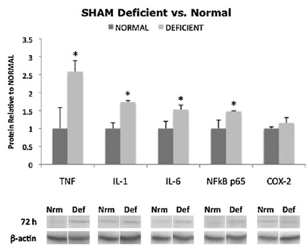
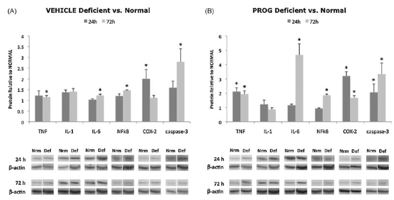
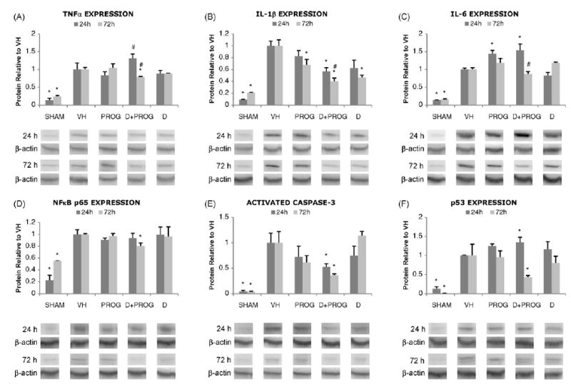
Administration of the neurosteroid progesterone (PROG) has been shown to be beneficial in a number of brain injury models and in two recent clinical trials. Given widespread vitamin D deficiency and increasing traumatic brain injuries (TBIs) in the elderly, we investigated the interaction of vitamin D deficiency and PROG with cortical contusion injury in aged rats. Vitamin D deficient (VitD-deficient) animals showed elevated inflammatory proteins (TNFα, IL-1β, IL-6, NFκB p65) in the brain even without injury. VitD-deficient rats with TBI, whether given PROG or vehicle, showed increased inflammation and greater open-field behavioral deficits compared to VitD-normal animals. Although PROG was beneficial in injured VitD-normal animals, in VitD-deficient subjects neurosteroid treatment conferred no improvement over vehicle. A supplemental dose of 1,25-dihydroxyvitamin D(3) (VDH) given with the first PROG treatment dramatically improved results in VitD-deficient rats, but treatment with VDH alone did not. Our results suggest that VitD-deficiency can increase baseline brain inflammation, exacerbate the effects of TBI, and attenuate the benefits of PROG treatment; these effects may be reversed if the deficiency is corrected.
Copyright © 2009 Elsevier Inc. All rights reserved.
Figures

References
-
- Asakura H, Aoshima K, Suga Y, Yamazaki M, Morishita E, Saito M, Miyamoto K, Nakao S. Beneficial effect of the active form of vitamin D3 against LPS-induced DIC but not against tissue-factor-induced DIC in rat models. Thromb Haemost. 2001;85:287–290. - PubMed
-
- Banerjee P, Chatterjee M. Antiproliferative role of vitamin D and its analogs—a brief overview. Mol Cell Biochem. 2003;253:247–254. - PubMed
-
- Barrera D, Avila E, Hernandez G, Halhali A, Biruete B, Larrea F, Diaz L. Estradiol and progesterone synthesis in human placenta is stimulated by calcitriol. J Steroid Biochem Mol Biol. 2007;103:529–532. - PubMed
-
- Bauer B, Hartz AM, Fricker G, Miller DS. Pregnane X receptor up-regulation of P-glycoprotein expression and transport function at the blood–brain barrier. Mol Pharmacol. 2004;66:413–419. - PubMed
Publication types
MeSH terms
Substances
Grants and funding
LinkOut - more resources
Full Text Sources
Medical
