. 2009 Sep 1;18(17):3178-93.
doi: 10.1093/hmg/ddp256. Epub 2009 May 29.

DNMT3B interacts with constitutive centromere protein CENP-C to modulate DNA methylation and the histone code at centromeric regions

Affiliations

DNMT3B interacts with constitutive centromere protein CENP-C to modulate DNA methylation and the histone code at centromeric regions

Suhasni Gopalakrishnan et al. Hum Mol Genet. .

Abstract

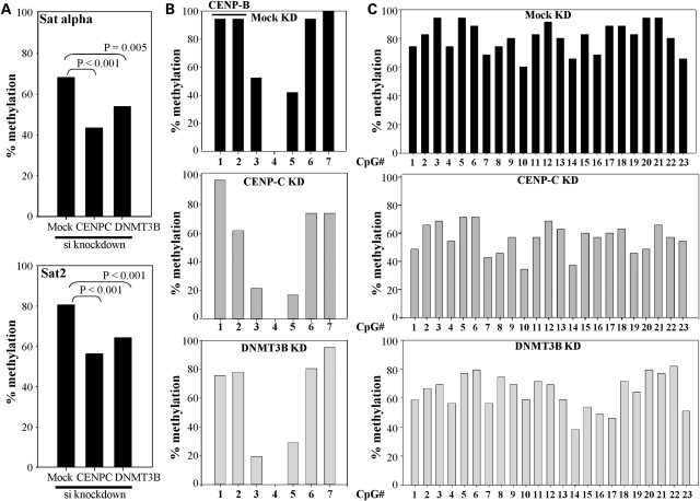
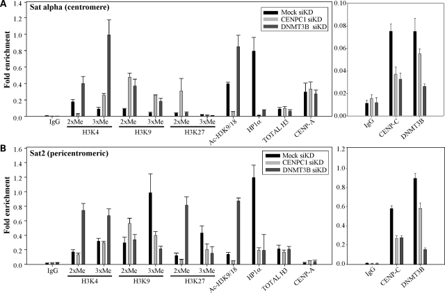
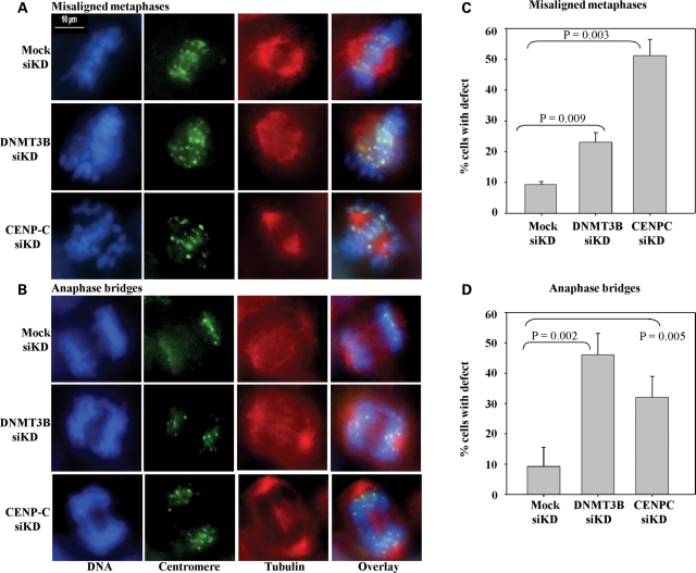
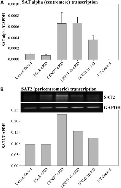
DNA methylation is an epigenetically imposed mark of transcriptional repression that is essential for maintenance of chromatin structure and genomic stability. Genome-wide methylation patterns are mediated by the combined action of three DNA methyltransferases: DNMT1, DNMT3A and DNMT3B. Compelling links exist between DNMT3B and chromosome stability as emphasized by the mitotic defects that are a hallmark of ICF syndrome, a disease arising from germline mutations in DNMT3B. Centromeric and pericentromeric regions are essential for chromosome condensation and the fidelity of segregation. Centromere regions contain distinct epigenetic marks, including dense DNA hypermethylation, yet the mechanisms by which DNA methylation is targeted to these regions remains largely unknown. In the present study, we used a yeast two-hybrid screen and identified a novel interaction between DNMT3B and constitutive centromere protein CENP-C. CENP-C is itself essential for mitosis. We confirm this interaction in mammalian cells and map the domains responsible. Using siRNA knock downs, bisulfite genomic sequencing and ChIP, we demonstrate for the first time that CENP-C recruits DNA methylation and DNMT3B to both centromeric and pericentromeric satellite repeats and that CENP-C and DNMT3B regulate the histone code in these regions, including marks characteristic of centromeric chromatin. Finally, we demonstrate that loss of CENP-C or DNMT3B leads to elevated chromosome misalignment and segregation defects during mitosis and increased transcription of centromeric repeats. Taken together, our data reveal a novel mechanism by which DNA methylation is targeted to discrete regions of the genome and contributes to chromosomal stability.


Figures

Figure 1.
Figure 1.
Identification of constitutive centromere protein CENP-C as a DNMT3B interacting protein in a yeast two-hybrid screen. (A) Summary of yeast two-hybrid results. Full-length human or murine DNMT3B1 fused to the GAL4-DBD interacts with the partial CENP-C clone (amino acids 200–943) isolated from the human testis cDNA library. Positive (p53 + TAg and DNMT3B self) and negative (p53 + Lamin and DNMT3B + empty GAL4-AD vector) controls for interaction are also shown. Results are presented as β-galactosidase activity units. (B) Mapping the CENP-C interacting region on DNMT3B. The murine Dnmt3b1 deletion constructs were fused to the GAL4-DBD and co-transfected into yeast strain Y190 with CENP-C (amino acids 23–943) fused to the GAL4-AD. Numbering refers to the amino acids of full-length murine Dnmt3b1. (C) Mapping the DNMT3B interacting region on CENP-C. As in (B), the indicated human CENP-C deletion constructs (fused to the GAL4-AD) were co-transfected into Y190 with full-length human DNMT3B1 (fused to the GAL4-DBD). Boxed regions in (B and C) indicate minimal interaction domains. ‘CBD’-centromere binding domain, roman numerals—conserved catalytic DNMT motifs.
Figure 2.
Figure 2.
DNMT3B co-immunoprecipitates (co-IPs) with CENP-C and CENP-A in mammalian cells. (A) Ectopically expressed DNMT3B and CENP-C interact in mammalian cells. The indicated constructs were transiently transfected into 293T cells then whole cell extract was prepared. The immunoprecipitating antibody (IP Ab) is indicated along the top of the western panel and the antibody used in western blotting (WB) is shown below it. Reciprocal co-IPs were performed. (B) Endogenous DNMT3B and CENP-C co-immunoprecipitate from HeLa nuclear extract (M phase). In the left panel, two different CENP-C antibodies (labeled 1 and 2) are used in the co-IP. In the right panel, the positive control of CENP-C interacting with CENP-A is also shown. (C) Confirmation that DNMT3B is a centromere-associated protein as shown by its ability to co-IP with the centromere-specific H3 variant CENP-A. The indicated constructs were co-transfected into 293T cells and used for reciprocal co-IP. Input—whole cell (A and C) or nuclear extract (B) prior to co-IP, IgG—negative control co-IP with species matched normal IgG.
Figure 3.
Figure 3.
DNMT3B interacts with two regions of CENP-C in mammalian cells. (A) Schematic representation of CENP-C with the deletion constructs used in the mapping studies indicated below. Numbering corresponds to amino acids of human CENP-C. Regions 1 and 2 are the minimal DNMT3B interacting regions mapped in (B). (B) Refining the DNMT3B interacting regions on CENP-C by co-immunoprecipitation. The CENP-C constructs indicated along the top of the western panel, fused to the HA tag, were co-transfected with full-length FLAG-tagged DNMT3B1 into 293T cells, whole cell extracts were prepared, and co-IP's were carried out with FLAG-agarose beads. The interaction between CENP-C and DNMT3B is lost when CENP-C regions 426–537 (region 1) and 638–760 (region 2) are deleted. (C) Inputs for the co-IP reactions in (B). The CENP-C constructs (denoted with arrows) were detected in whole cell extract of transfected 293T cells with HA antibody. The near full-length CENP-C (23–943) was run on a separate, lower percentage gel.
Figure 4.
Figure 4.
A fraction of DNMT3B co-localizes with CENP-C particularly during metaphase. HeLa cells were transfected with GFP-tagged DNMT3B1 (green panels), synchronized with a double thymidine block, released, then fixed when cells were in mitosis. Interphase cells were also examined. Transfected cells were stained with anti-CENP-C (red panels) antibody. DNA was stained with DAPI (blue panels). An overlay of the red and green channels is shown in the right-most panels. Representative images of cells in interphase (top row) or the different phases of mitosis (lower four rows) are shown. Select regions are enlarged in the small red-boxed regions of the overlay panel to highlight closely opposed or overlapping DNMT3B and CENP-C foci. Double red arrowheads indicate closely juxtaposed foci. Scale bar—10 µm.
Figure 5.
Figure 5.
CENP-C modulates DNA methylation at centromeric and pericentric regions. (A) Summary of bisulfite genomic sequencing (BGS) results, as the total percent methylation at all CpG sites examined in the alpha satellite (top) or pericentric satellite 2 (bottom) regions in HCT116 cells mock transfected or transfected with siRNA directed against CENP-C or DNMT3B. Statistical significance was determined using the χ2-test. For the alpha satellite region, BGS summary data is derived from 38 mock, 42 CENP-C and 41 DNMT3B siRNA knock down clones. For satellite 2, summary data is derived from 35 mock, 35 CENP-C and 39 DNMT3B siRNA knock down clones. (B) BGS analysis of the seven CpG sites within the alpha satellite region in mock or siRNA targeted HCT116 cells. Sites 1 and 2 are within the CENP-B binding region as indicated on the graph. The total percent methylation at each CpG site is summarized and derived from data in Supplementary Material, Figure S3. (C) BGS analysis of the satellite 2 region containing 23 CpG sites. Data is summarized as in (B) and is derived from data in Supplementary Material, Figure S4.
Figure 6.
Figure 6.
CENP-C and DNMT3B modulate epigenetic marks at the centromeric and pericentromeric regions. Chromatin immunoprecipitation (ChIP) and quantitative PCR were used to evaluate the effect of CENP-C (light gray bars) or DNMT3B (dark gray bars) siRNA knock down in HCT116 cells, relative to a mock transfection (black bars). (A) ChIP for the indicated histone marks, HP1α and histone H3/CENP-A (left graph) or DNMT3B and CENP-C (right panel) followed by PCR for alpha satellite DNA. (B) ChIP with the same panel of antibodies as in (A) followed PCR for satellite 2 sequences. Results are presented as the average fold-enrichment relative to the input (1% of the supernatant from the IgG ChIP reaction). All reactions were repeated at least in triplicate from two independent siRNA knock downs. The error bar denotes the standard deviation from the mean. A similar analysis of control single copy genes (WIF1 and GAPDH) is shown in Supplementary Material, Figure S6. The scale in the right panels was expanded as the ChIP signal for CENP-C and DNMT3B was generally lower than that of the histone marks. IgG—negative control ChIP with normal rabbit IgG to determine background binding, 2XMe, 3XMe—di- and trimethylated forms, Ac—acetylation.
Figure 7.
Figure 7.
Disrupting the CENP-C–DNMT3B interaction results in enhanced mitotic defects. HCT116 cells were transfected with siRNA against CENP-C or DNMT3B or were mock transfected. Following transfection, cells were synchronized, released and then fixed during M phase. Cells were then stained for DNA (blue panels), the centromere (anti-centromere antibody, green) and the mitotic spindle with anti-tubulin antibody (red). Mock transfected cells showed few mitotic defects, whereas cells with reduced levels of CENP-C or DNMT3B demonstrated elevated mitotic defects (misaligned chromosomes and anaphase bridges). (A and B) Representative images of cells undergoing normal or defective mitoses. (C and D) Quantification of mitotic defects in mock or siRNA knock down cells. At least 50 mitotic cells were counted from three independent siRNA knock downs. Statistical significance was evaluated using the Students t-test. Scale bar—10 µm.
Figure 8.
Figure 8.
Loss of CENP-C and DNMT3B-mediated epigenetic marks at the centromere region result in elevated levels of repeat transcription. (A) Quantitative RT–PCR analysis of alpha satellite repeat transcripts in HCT116 cells either mock transfected or transfected with siRNA targeting CENP-C or DNMT3B. HCT116 cells with a genetic knockout of DNMT3B (3BKO) are also shown. Values are the average of triplicate RT–PCR reactions relative to GAPDH as a loading control. The error bar is the standard deviation from the mean. (B) Semi-quantitative RT–PCR analysis of satellite 2 repeat transcription from the same knock down/knockout panel as in (A). RT–PCR reactions were repeated three times and a representative ethidium bromide stained agarose gel photo is shown. Bands were quantified using BioRad Quantity One software and set relative to an independent amplification for GAPDH (lower panel). Although samples were DNase treated, we included a no RT (-RT) control to ensure that we were not amplifying contaminating satellite DNA in the RNA preparation.

Similar articles

Cited by

References

    1. Goll M.G., Bestor T.H. Eukaryotic cytosine methyltransferases. Annu. Rev. Biochem. 2005;74:481–514. - PubMed
    1. Li E. Chromatin modification and epigenetic reprogramming in mammalian development. Nature Rev. Genet. 2002;3:662–673. - PubMed
    1. Robertson K.D. DNA methylation and human disease. Nature Rev. Genet. 2005;6:597–610. - PubMed
    1. Jones P.A., Baylin S.B. The fundamental role of epigenetic events in cancer. Nature Rev. Genet. 2002;3:415–428. - PubMed
    1. Berger S.L. The complex language of chromatin regulation during transcription. Nature. 2007;447:407–412. - PubMed

Publication types

Substances