. 2010 Feb;20(2):304-14.
doi: 10.1093/cercor/bhp100. Epub 2009 May 29.

Tis21 expression marks not only populations of neurogenic precursor cells but also new postmitotic neurons in adult hippocampal neurogenesis

Affiliations

Tis21 expression marks not only populations of neurogenic precursor cells but also new postmitotic neurons in adult hippocampal neurogenesis

Alessio Attardo et al. Cereb Cortex. 2010 Feb.

Abstract

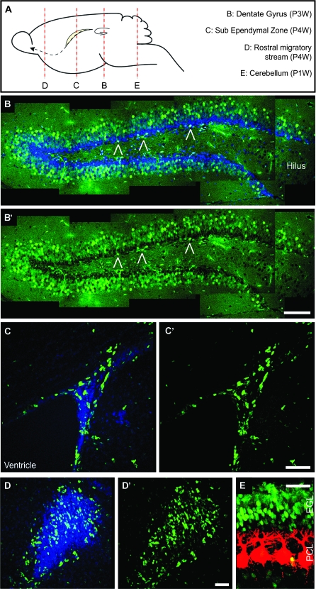
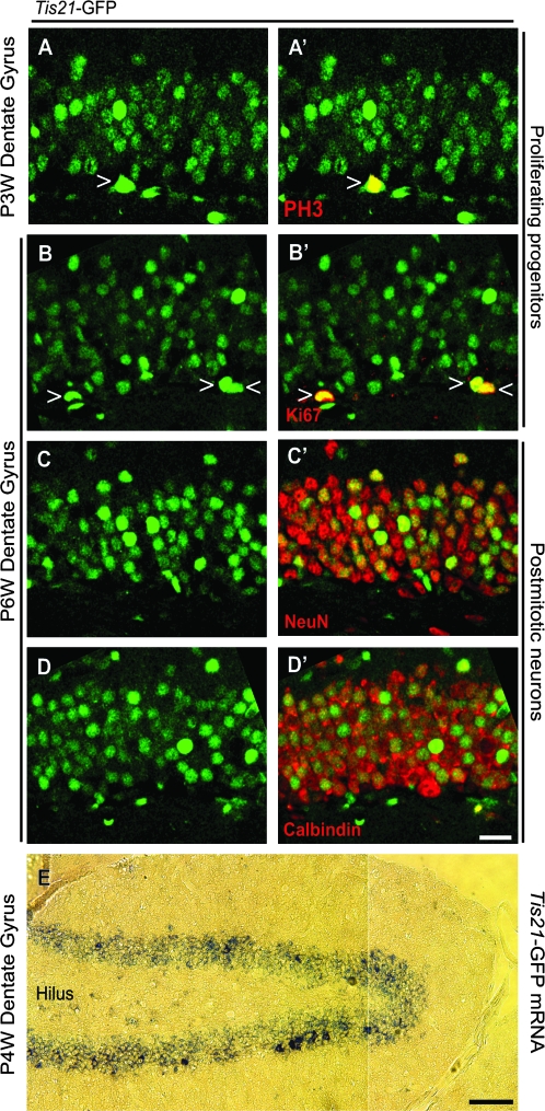
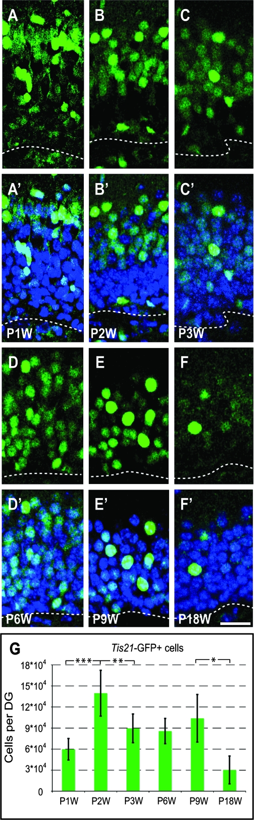
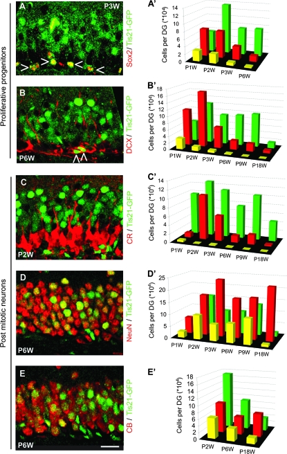
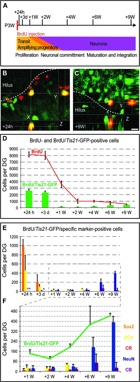
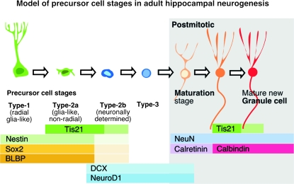
During embryonic cortical development, expression of Tis21 is associated with cell cycle lengthening and neurogenic divisions of progenitor cells. We here investigated if the expression pattern of Tis21 also correlates with the generation of new neurons in the adult hippocampus. We used Tis21 knock-in mice expressing green fluorescent protein (GFP) and studied Tis21-GFP expression together with markers of adult hippocampal neurogenesis in newly generated cells. We found that Tis21-GFP 1) was absent from the radial glia-like putative stem cells (type-1 cells), 2) first appeared in transient amplifying progenitor cells (type-2 and 3 cells), 3) did not colocalize with markers of early postmitotic maturation stage, 4) was expressed again in maturing neurons, and 5) finally decreased in mature granule cells. Our data show that, in the course of adult neurogenesis, Tis21 is expressed in a phase additional to the one of the embryonic neurogenesis. This additional phase of expression might be associated with a new and different function of Tis21 than during embryonic brain development, where no Tis21 is expressed in mature neurons. We hypothesize that this function is related to the final functional integration of the newborn neurons. Tis21 can thus serve as new marker for key stages of adult neurogenesis.


Figures

Figure 1.
Figure 1.
Intrinsic Tis21-GFP fluorescence in the postnatal and adult mouse brain. (A) Diagram showing the main postnatal neurogenic regions and indicating the position of the sections in (BE). (BE) Confocal scanning photomicrographs (single optical sections, 1 μm each) of 40-μm sections through the P3W DG (B and B′), P4W SVZ (CD′), and P1W cerebellum (E) of Tis21-GFP mice showing intrinsic Tis21-GFP fluorescence (green) and CB (E, red) immunofluorescence; blue, Hoechst staining of nuclei (B, C, and D). (B and B′) are composite pictures including different optical fields. Open triangles indicate Tis21-GFP–positive cells in the SGZ. EGL, external GCL; PCL, Purkinje cell layer. Scale bars: (B′), 100 μm; (C′), 50 μm; (D′), 40 μm; (E), 30 μm.
Figure 2.
Figure 2.
Tis21-GFP is expressed by mitotic progenitor cells as well as postmitotic granule cells. (AD′) Confocal scanning photomicrographs (single optical sections, 1 μm each) of 40-μm sections through the P3W (A and A′) and P6W (BD′) DG of Tis21-GFP mice showing intrinsic Tis21-GFP fluorescence (green) and its colocalization with PH3 (A′ red, open triangle), Ki67 (B′ red, open triangles), NeuN (C′ red), and CB (D′ red) immunofluorescence. (E) Bright-field photomicrograph of 10-μm cryosection through the P4W DG of Tis21-GFP mice showing in situ hybridization for Tis21-GFP mRNA (blue). The images in (AD′) are oriented with the hilus at the bottom. Scale bars: (D′), 20 μm; (E), 100 μm.
Figure 3.
Figure 3.
Tis21-GFP–positive cells during formation and maturation of the DG. (AF′) Confocal scanning photomicrographs (single optical sections, 1 μm each) of 40-μm sections through the P1W (A and A′), P2W (B and B′), P3W(C and C′), P6W (D and D′), P9W (E and E′), and P18W (F and F′) DG of Tis21-GFP mice showing intrinsic Tis21-GFP fluorescence (green); blue, Hoechst staining of nuclei. Dashed lines, boundary to the hilus. Scale bar: (F′), 20 μm. (G) Tis21-GFP–positive cells (intrinsic fluorescence) per entire DG at various time points of postnatal development. Data are the mean of 3–12 animals; error bars indicate standard deviations. Triple asterisk, P < 0.001; double asterisk, P < 0.01; single asterisk, P < 0.02.
Figure 4.
Figure 4.
Tis21-GFP expression in precursor cells and postmitotic neurons during formation and maturation of the DG. (AE) Confocal scanning photomicrographs (single optical sections, 1 μm each) of 40-μm sections through the P3W (A), P6W (B, D, E), and P2W (C) DG of Tis21-GFP mice showing intrinsic Tis21-GFP fluorescence (green) and Sox2 (A), Dcx (B), CR (C), NeuN (D), and CB (E) immunofluorescence (red). Open triangles indicate Tis21-GFP–positive cells that are immunoreactive for Sox2 (A) or Dcx (B). Scale bar: (E), 20 μm. (A′E′) Green bars indicate the number of Tis21-GFP–positive cells; red bars the number of Sox2- (A′), Dcx- (B′), CR- (C′), NeuN- (D′), and CB (E′)-positive cells and yellow bars the number of cells double positive for Tis21-GFP and the respective marker, in each case per entire DG. Data are the mean of 2–10 animals; here no error bars are shown for clarity.
Figure 5.
Figure 5.
BrdU pulse chase for Tis21-GFP cells. (A) Schematic drawing showing the pulse-chase strategy in relation to postnatal DG development: 3-week-old Tis21-GFP heterozygous mice were injected with BrdU (50 mg/kg) twice, with a 3-h interval between injections. Brains were collected 24 h, 3 days, and 1, 2, 4, 6, and 9 weeks after injection. (BC) Confocal scanning photomicrographs and stacks of 7 (B) and 6 (C) single optical sections (1 μm each) of 40-μm-thick sections through the P3W + 24 h (B) and P3W + 9 weeks (C) DG of Tis21-GFP mice showing Tis21-GFP (green) and BrdU (red) double immunofluorescence. Dashed lines, boundary to the hilus. Scale bar in (C), 10 μm. (D) Number of total BrdU-positive cells (red curve) and the BrdU-positive cells showing Tis21-GFP immunofluorescence (green bars) per entire DG at various time points after the BrdU pulse. Data are the mean of 3 animals; error bars indicate standard deviations. (EF) Number of BrdU/Tis21-GFP–double-positive cells showing Sox2 (orange), Dcx (yellow), CR (red), NeuN (blue), and the CB (purple) immunoreactivity per entire DG at various time points after the BrdU pulse. Number of BrdU/Tis21-GFP–double-positive cells (green curve) compared with BrdU/Tis21-GFP–double-positive cells showing Sox2 (orange bars), Dcx (yellow bars), CR (red bars), NeuN (blue bars), and CB (purple bars) immunoreactivity per entire DG at selected time points after the BrdU pulse. Data are the mean of 2 or 3 animals; bars indicate standard deviation in all cases in which 3 animals were analyzed and standard error at P24h and P9W for CB where 2 animals were examined. Asterisks in the respective colors indicate that no positive cell could be detected (but the analysis was done).
Figure 6.
Figure 6.
Tis21-GFP expression in the course of adult hippocampal neurogenesis. Simplified schematic drawing showing a model of adult hippocampal neurogenesis and Tis21-GFP expression in relation to expression of other markers along the cell lineage of the DG.

Similar articles

Cited by

References

    1. Altman J, Bayer SA. Migration and distribution of two populations of hippocampal granule cell precursors during the perinatal and postnatal periods. J Comp Neurol. 1990a;301:365–381. - PubMed
    1. Altman J, Bayer SA. Vertical compartmentation and cellular transformations in the germinal matrices of the embryonic rat cerebral cortex. Exp Neurol. 1990b;107:23–35. - PubMed
    1. Attardo A, Calegari F, Haubensak W, Wilsch-Brauninger M, Huttner WB. Live imaging at the onset of cortical neurogenesis reveals differential appearance of the neuronal phenotype in apical versus basal progenitor progeny. PLoS ONE. 2008;3:e2388. - PMC - PubMed
    1. Brandt MD, Jessberger S, Steiner B, Kronenberg G, Reuter K, Bick-Sander A, von der Behrens W, Kempermann G. Transient calretinin expression defines early postmitotic step of neuronal differentiation in adult hippocampal neurogenesis of mice. Mol Cell Neurosci. 2003;24:603–613. - PubMed
    1. Breunig JJ, Silbereis J, Vaccarino FM, Sestan N, Rakic P. Notch regulates cell fate and dendrite morphology of newborn neurons in the postnatal dentate gyrus. Proc Natl Acad Sci USA. 2007 Dec 18;104(51):20558–20563. - PMC - PubMed

Publication types

MeSH terms