. 2009 Sep 1;18(17):3227-43.
doi: 10.1093/hmg/ddp261. Epub 2009 May 30.

Serine racemase is associated with schizophrenia susceptibility in humans and in a mouse model

Affiliations

Serine racemase is associated with schizophrenia susceptibility in humans and in a mouse model

Viviane Labrie et al. Hum Mol Genet. .

Abstract

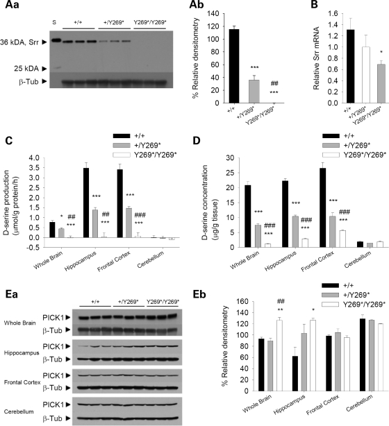
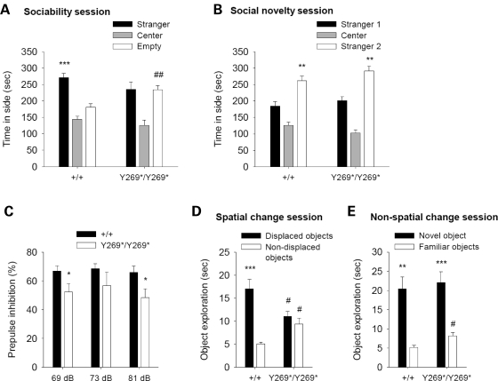
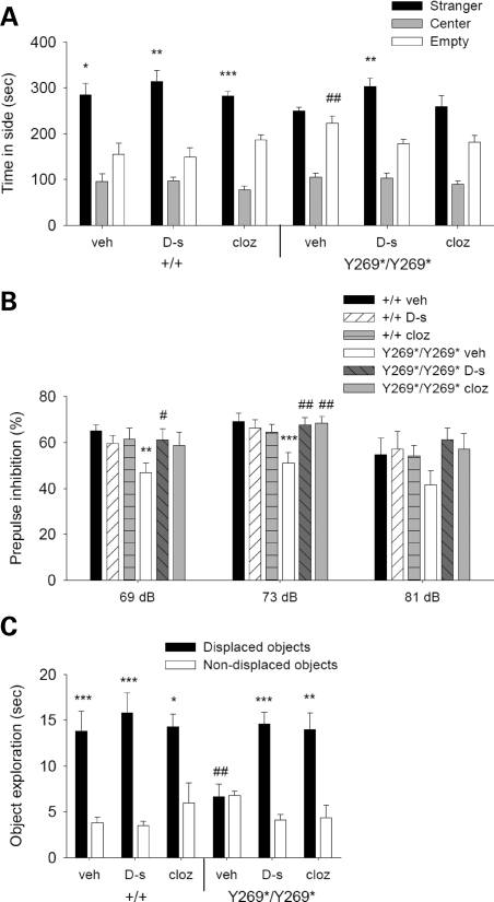
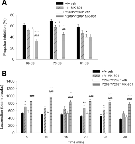
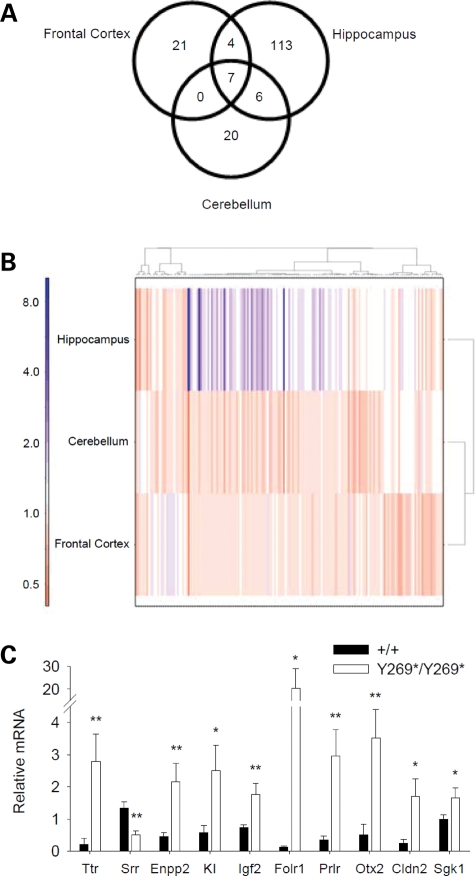
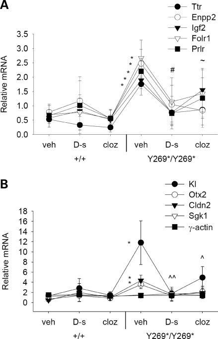
Abnormal N-methyl-d-aspartate receptor (NMDAR) function has been implicated in the pathophysiology of schizophrenia. d-serine is an important NMDAR modulator, and to elucidate the role of the d-serine synthesis enzyme serine racemase (Srr) in schizophrenia, we identified and characterized mice with an ENU-induced mutation that results in a complete loss of Srr activity and dramatically reduced d-serine levels. Mutant mice displayed behaviors relevant to schizophrenia, including impairments in prepulse inhibition, sociability and spatial discrimination. Behavioral deficits were exacerbated by an NMDAR antagonist and ameliorated by d-serine or the atypical antipsychotic clozapine. Expression profiling revealed that the Srr mutation influenced several genes that have been linked to schizophrenia and cognitive ability. Transcript levels altered by the Srr mutation were also normalized by d-serine or clozapine treatment. Furthermore, analysis of SRR genetic variants in humans identified a robust association with schizophrenia. This study demonstrates that aberrant Srr function and diminished d-serine may contribute to schizophrenia pathogenesis.


Figures

Figure 1.
Figure 1.
Biochemical changes in mice with a nonsense mutation in exon 9 of Srr. (Aa) Western blot examining Srr levels using a monoclonal Srr antibody to probe protein extracts from whole brain of wild-type (+/+), heterozygous (+/Y269*) and mutant (Y269*/Y269*) mice. A loss of Srr protein was observed in mutant mice. β-Tubulin III was used as a loading control. The molecular weights in kiloDaltons (kDa) of Srr and a protein standard (S) are marked. (Ab) Densitometric quantification of western blot for Srr. The mean densitometry (± SEM) is an indicator of Srr protein levels in wild-type, heterozygote and mutant animals (genotype, F2,6 = 140.9, P < 0.001). ***P < 0.001 compared with wild-type mice; ##P < 0.01 compared with heterozygous mice. (B) Real-time RT–PCR analysis of Srr mRNA. Mean levels of Srr mRNA (± SEM) assessed in whole brain of wild-type (n = 6), heterozygous (n = 7) and mutant mice (n = 6) revealed a significantly reduced abundance of Srr mRNA in mutant animals. *P < 0.05 compared with wild-type mice. (C) In an Srr activity assay, the mean d-serine production (± SEM) by endogenous Srr was examined in the whole brain, hippocampus, frontal cortex and cerebellum of wild-type (n = 7–8), heterozygous (n = 4–6) and mutant mice (n = 5–7). An absence of Srr activity was found in mutant animals. *P < 0.05, ***P < 0.001 compared with wild-type mice, within the same brain region; ##P < 0.01, ###P < 0.001 compared with heterozygous mice, within the same brain region. (D) Mean concentration (± SEM) of d-serine was examined by HPLC in the whole brain, hippocampus, frontal cortex and cerebellum of wild-type (n = 5–8), heterozygous (n = 6–9) and mutant mice (n = 6–8). Lower d-serine concentrations were found in the whole brain, hippocampus, frontal cortex, but not the cerebellum of mutant mice. ***P < 0.001 compared with wild-type mice, within the same brain region; ###P < 0.001 compared with heterozygous mice, within the same brain region. (Ea) Western blot assessing PICK1 protein levels in whole brain, hippocampus, frontal cortex and cerebellum of wild-type, heterozygous and mutant mice. PICK1 was found to be elevated in the whole brain and hippocampus of mutant animals. β-Tubulin III was used as a loading control. (Eb) Densitometric quantification of western blot for PICK1. The mean densitometry (± SEM) indicates PICK1 protein levels in wild-type, heterozygote and mutant animals (genotype, F2,6 = 10.7, P < 0.05; genotype×brain region interaction, F6,18 = 5.6, P < 0.01). *P < 0.05, **P < 0.01 compared with wild-type mice, within the same brain region; ##P < 0.01 compared with heterozygous mice, within the same brain region.
Figure 2.
Figure 2.
Schizophrenia-like behaviors in SrrY269* mutant mice. (A) Sociability session in social affiliations task. Mean time spent in a chamber containing a stranger mouse, a central chamber and a chamber with an empty cage. In contrast to wild-type mice (+/+; n = 13), mutant animals (Y269*/Y269*; n = 12) did not favor the chamber with the stranger mouse (genotype×apparatus side, F1,23 = 6.7, P < 0.05). ***P < 0.001 compared with the chamber with empty cage, within genotype; ##P < 0.01 compared with wild-type mice in the same chamber. (B) Social novelty session in social affiliations task. Mean time spent in a chamber containing a newly introduced mouse (stranger 2), a central chamber and a chamber with a familiar mouse (stranger 1). Both wild-type and mutant mice preferred the chamber with the novel stranger (apparatus side, F1,23=26.2, P < 0.001). **P < 0.01 compared with the chamber with stranger 1, within each genotype. (C) Prepulse inhibition (PPI) of the acoustic startle response. Mean % inhibition of the startle response at prepulse intensities of 69, 73 and 81 dB. Compared with wild-type animals (n = 13), mutant mice (n = 12) demonstrated a PPI deficit (genotype, F1,23 = 6.9, P < 0.05; genotype with collapsed prepulse intensities P < 0.02). *P < 0.05 compared with wild-type mice, within the same prepulse intensity. (D) Spatial change session in the object recognition task. Mean time spent exploring the displaced and non-displaced objects. Wild-type (n = 8), but not mutant mice (n = 11), showed greater exploration of the objects that underwent a spatial change (object category, F1,17 = 65.6, P < 0.001; genotype×object category, F1,17 = 36.7, P < 0.001). ***P < 0.001 compared with the non-displaced objects, within genotype; #P < 0.05 compared with wild-type mice exploring the same object category. (E) Non-spatial change session in the object recognition task. Mean time spent exploring the novel item and three familiar objects. Both wild-type and mutant mice preferentially explored the novel object (object category, F1,17 = 52.4, P < 0.001). **P < 0.01, ***P < 0.001 compared with the familiar objects, within each genotype; #P < 0.05 compared with wild-type mice exploring the same object category. Data are shown as mean ± SEM.
Figure 3.
Figure 3.
d-serine (600 mg/kg) and clozapine (0.75 mg/kg) improve schizophrenia-like behaviors in mutant mice. (A) Mean time in a chamber containing a stranger mouse, a central chamber and a chamber with an empty cage during a test of sociability. Wild-type animals (+/+) given vehicle (veh; n = 10), d-serine (d-s; n = 8) or clozapine (cloz; n = 10) and mutant mice (Y269*/Y269*) treated with d-serine (n = 9) significantly preferred the chamber with the unfamiliar mouse, whereas mutant mice injected with vehicle (n = 10) or clozapine (n = 11) did not (genotype×apparatus side, F1,52 = 4.3, P < 0.05). *P < 0.05, **P < 0.01, ***P < 0.001 compared with the chamber with empty cage, within genotype and drug treatment group; ##P < 0.01 compared with vehicle-treated wild-type mice in the same chamber. (B) Mean % reduction of startle amplitude at prepulse intensities of 69, 73 and 81 dB. The PPI deficit in vehicle-treated mutant mice (n = 11) was improved by d-serine (n = 12) and clozapine (n = 8) to a level comparable to wild-type animals treated with vehicle (n = 9), d-serine (n = 9) or clozapine (n = 9) (genotype×drug treatment, F2,52 = 4.3, P < 0.05). **P < 0.01, ***P < 0.001 compared with vehicle-treated wild-type mice, within the same prepulse intensity; #P < 0.05, ##P < 0.01 compared with vehicle-treated mutant mice, within the same prepulse intensity. (C) Mean time spent investigating displaced and non-displaced objects in the spatial recognition task. Rescue of the impaired spatial reactivity in vehicle-treated mutant mice (n = 10) was apparent in mutant animals treated with d-serine (n = 8) and clozapine (n = 9), which demonstrated similar responses to wild-type mice given vehicle (n = 9), d-serine (n = 9) or clozapine (n = 10) (genotype×drug treatment×object category, F2,49 = 4.3, P < 0.05). *P < 0.05, **P < 0.01, ***P < 0.001 compared with the non-displaced objects, within genotype and drug treatment group; ##P < 0.01 compared with vehicle-treated wild-type mice exploring the same object category. Data are presented as mean ± SEM.
Figure 4.
Figure 4.
Potentiation of behavioral responses relevant to schizophrenia following NMDAR inhibition. (A) Mean % inhibition of the acoustic startle response with prepulse intensities of 69, 73 and 81 dB. Wild-type (+/+) and mutant animals (Y269*/Y269*) were treated with vehicle (n = 17, 24) or MK-801 (0.1 mg/kg; n = 16, 17). PPI in mutant mice given the NMDAR antagonist was most severely affected (genotype, F1,70 = 18.8, P<0.001; drug treatment, F1,70 = 15.6, P < 0.001; genotype×drug treatment×prepulse intensity, F2,140 = 3.0, P ≤ 0.05). *P < 0.05 compared with vehicle-treated wild-type mice, within the same prepulse intensity; ##P < 0.01, ###P < 0.001 compared with vehicle-treated mutant mice, within the same prepulse intensity; ∼P < 0.05, ∼∼∼P < 0.001 compared with MK-801-treated wild-type mice, within the same prepulse intensity. (B) Mean number of beam breaks in an empty open field during a 30-min assessment of locomotor activity (5-min bins). Wild-type and mutant animals were given vehicle (n = 9, 9) or MK-801 (0.1 mg/kg; n = 10, 9), and locomotor activity was most enhanced in mutant mice administered the NMDAR antagonist (genotype, F1,33 = 9.5, P < 0.01; drug treatment, F1,33 = 35.2, P < 0.001; genotype×drug treatment, F1,33 = 4.0, P ≤ 0.05). *P < 0.05, **P < 0.01 compared with vehicle-treated wild-type mice, within the same time bin; ###P < 0.001 compared with vehicle-treated mutant mice, within the same time bin; ∼P < 0.05, ∼∼P < 0.01, ∼∼∼P < 0.001 compared with MK-801-treated wild-type mice, within the same time bin. Data are shown as mean ± SEM.
Figure 5.
Figure 5.
Changes in mRNA levels due to a loss of Srr function. (A) Venn diagram demonstrating the number of altered transcripts in response to the SrrY269* mutation and the overlapping transcripts in the hippocampus, frontal cortex and cerebellum. (B) Heatmap displaying the clustering patterns of all differentially expressed genes in the hippocampus, cerebellum and frontal cortex of SrrY269* mice. The color scale indicates the fold change. The hippocampus shows the greatest number and magnitude of changes. (C) Real-time RT–PCR analysis. In the hippocampus of mutant mice (Y269*/Y269*; n = 5) the mRNA levels of Ttr, Enpp2, KI, Igf2, Folr1, Prlr, Otx2, Cldn2 and Sgk1 were increased, while Srr mRNA levels were reduced relative to wild-type animals (+/+; n = 7) (genotype, F1,10 = 11.5, P < 0.01). *P < 0.05, **P < 0.01 compared with wild-type mice, within the same gene.
Figure 6.
Figure 6.
Differentially expressed mRNA transcripts in SrrY269* mice are normalized by d-serine or clozapine treatment. (A and B) Levels of mRNA in the hippocampus of wild-type (+/+) and mutant mice (Y269*/Y269*) were assessed 30 min following vehicle, d-serine (600 mg/kg) or clozapine (0.75 mg/kg) administration. Vehicle-treated mutant mice (n = 7) demonstrated an enhancement in Ttr, Enpp2, KI, Igf2, Folr1, Prlr, Otx2 and Cldn2 mRNA that was reversed by d-serine (n = 5) or clozapine (n = 5) treatment to a level similar to wild-type mice administered vehicle (n = 6), d-serine (n = 7) or clozapine (n = 7) (genotype, F1,30 = 4.5, P < 0.05; genotype×drug treatment×gene interaction, F16,240 = 2.6, P < 0.01). γ-actin was used as a negative control. *P < 0.05 compared with vehicle-treated wild-type mice, within the same gene; #P < 0.05 Igf2 mRNA levels in d-serine-treated mutant mice compared with vehicle-treated mutant mice; ∼P < 0.05 Enpp2 mRNA levels in clozapine-treated mutant mice compared with vehicle-treated mutant mice; ^P < 0.05; ^^P < 0.01 Kl mRNA levels in d-serine- or clozapine-treated mutant mice compared with vehicle-treated mutant mice.

References

    1. Lewis D.A., Gonzalez-Burgos G. Pathophysiologically based treatment interventions in schizophrenia. Nature Med. 2006;12:1016–1022. - PubMed
    1. Ross C.A., Margolis R.L., Reading S.A., Pletnikov M., Coyle J.T. Neurobiology of schizophrenia. Neuron. 2006;52:139–153. - PubMed
    1. Insel T.R., Scolnick E.M. Cure therapeutics and strategic prevention: raising the bar for mental health research. Mol. Psychiatry. 2006;11:11–17. - PMC - PubMed
    1. Coyle J.T. Glutamate and schizophrenia: beyond the dopamine hypothesis. Cell. Mol. Neurobiol. 2006;26:365–384. - PMC - PubMed
    1. Javitt D.C., Zukin S.R. Recent advances in the phencyclidine model of schizophrenia. Am. J. Psychiatry. 1991;148:1301–1308. - PubMed

Publication types