. 2009 Jul;41(7):843-8.
doi: 10.1038/ng.392. Epub 2009 May 31.

Lin28 promotes transformation and is associated with advanced human malignancies

Affiliations

Lin28 promotes transformation and is associated with advanced human malignancies

Srinivas R Viswanathan et al. Nat Genet. 2009 Jul.

Abstract

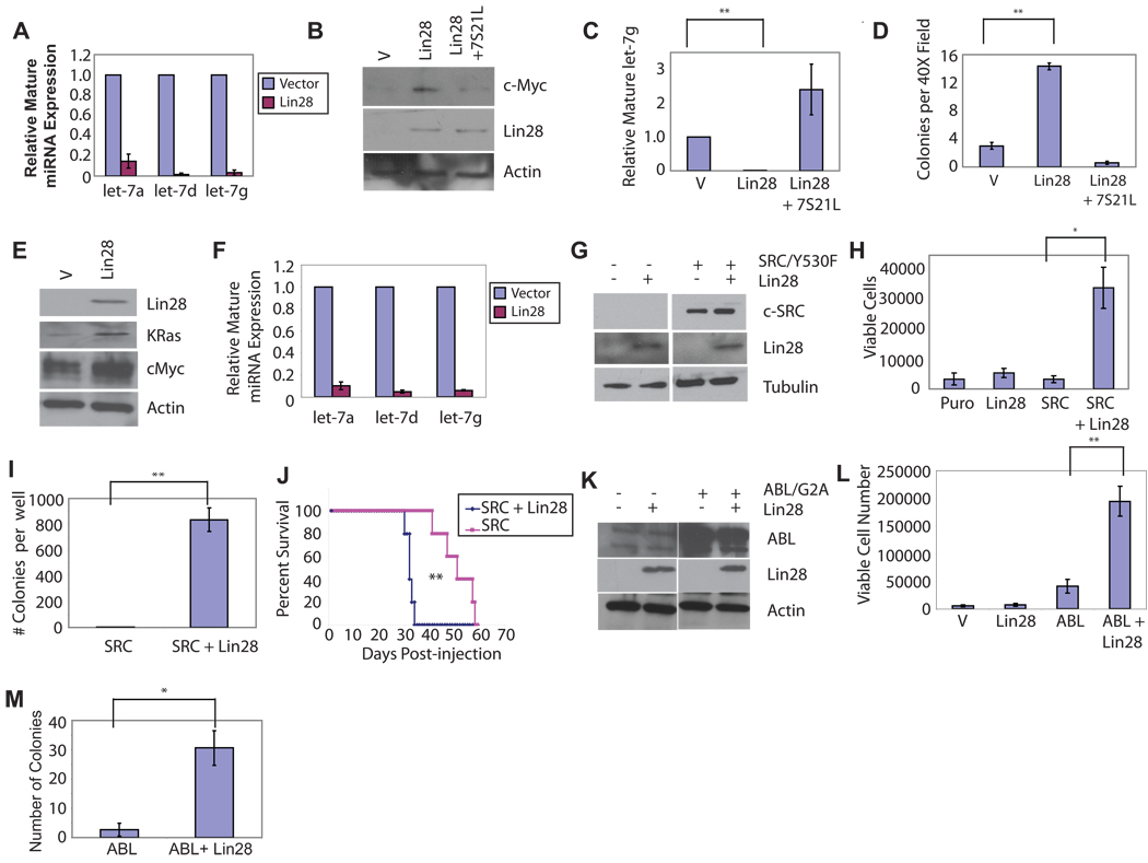
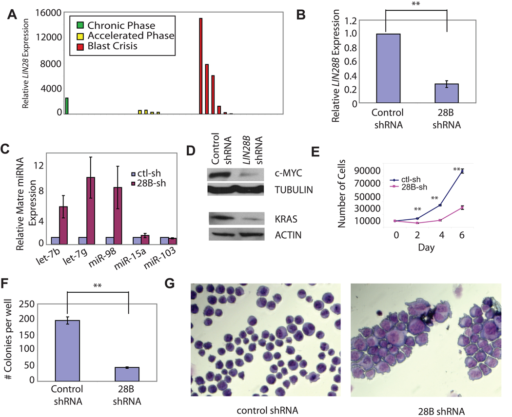
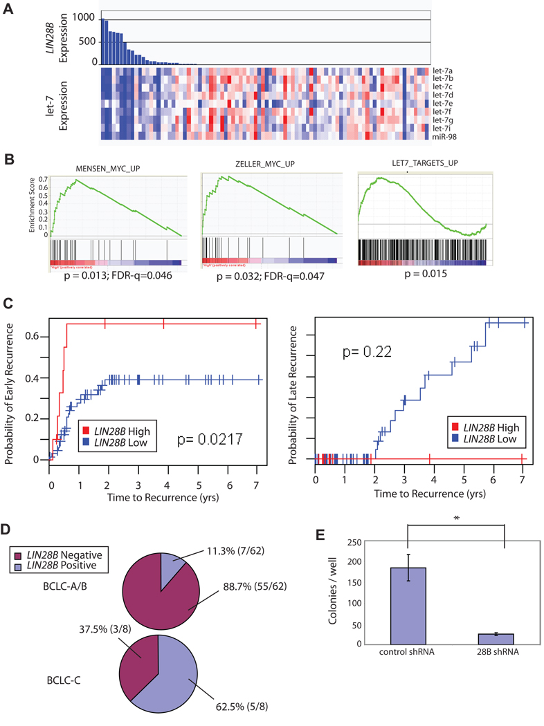
Multiple members of the let-7 family of miRNAs are often repressed in human cancers, thereby promoting oncogenesis by derepressing targets such as HMGA2, K-Ras and c-Myc. However, the mechanism by which let-7 miRNAs are coordinately repressed is unclear. The RNA-binding proteins LIN28 and LIN28B block let-7 precursors from being processed to mature miRNAs, suggesting that their overexpression might promote malignancy through repression of let-7. Here we show that LIN28 and LIN28B are overexpressed in primary human tumors and human cancer cell lines (overall frequency approximately 15%), and that overexpression is linked to repression of let-7 family miRNAs and derepression of let-7 targets. LIN28 and LIN28b facilitate cellular transformation in vitro, and overexpression is associated with advanced disease across multiple tumor types. Our work provides a mechanism for the coordinate repression of let-7 miRNAs observed in a subset of human cancers, and associates activation of LIN28 and LIN28B with poor clinical prognosis.


Figures

Figure 1
Figure 1. Lin28 Transforms NIH/3T3 cells
(A) Levels of mature miR species in representative infection determined by quantitative PCR in NIH/3T3 cells infected with pMSCV.Neo or pMSCV.Neo.Lin28, and selected on G418. (B) Western blot analysis of extracts from NIH/3T3 cells infected with MSCV.Neo/pBabe.Puro, pMSCV.Neo.Lin28/pBabe.Puro, or pMSCV.Neo.Lin28/pBabe.Puro.7S21L, selected with Puromycin and G418. (C) Levels of mature let-7g in NIH/3T3 cells infected with MSCV.Neo/pBabe.Puro,pMSCV.Neo.Lin28/pBabe.Puro, or pMSCV.Neo.Lin28/pBabe.Puro.7S21L, selected with Puromycin and G418. (D) Colony numbers in semisolid media for 25,000 NIH/3T3 cells infected with pMSCV.Neo/pBabe.Puro, pMSCV.Neo.Lin28/pBabe.Puro, or pMSCV.Neo.Lin28/pBabe.Puro.7S21L, selected with Puromycin and G418. Colonies were counted after 4 weeks with five random fields counted per well. Results are plotted as average colony number +/− S.E.M., N=3. (E) Western blot analysis on cell extracts from Ba/F3 cells infected with pMSCV.Neo and pMSCV.Neo.Lin28, selected on G418. (F) Levels of mature miR species in representative infection determined by quantitative PCR in Ba/F3 cells infected with pMSCV.Neo or pMSCV.Neo.Lin28, and selected on G418. (G) Western blot analysis on cell extracts from Ba/F3 cells infected with pEYK.Puro.SrcY530F and either pMSCV.Neo or pMSCV.Neo.Lin28 and selected on puromycin and G418 (H) Ba/F3 lines established in (C) were plated in media without IL-3 at 10,000 cells per well. Cells were stained with Trypan blue and viable cells were counted 6d after plating. (I) Colony formation of Ba/F3 cells expressing Src/Y530F or Src/Y530F + Lin28 in semisolid medium, plated at 50,000 cells per well in medium without IL-3. Quantitation of colony number from soft agar assay after 21d of growth. Results are plotted as average colony number per well +/− S.E.M., N=3. (J) Survival of immunocompromised mice injected with 107 Ba/F3 cells expressing Src-Y530F or Src-Y530F+Lin28. Average survival time 51.4d vs. 33.2d vs. respectively, p=0.00276 (K) Western blot analysis of extracts from Ba/F3 cells infected with pBabe.Puro or pBabe.Puro.Lin-28, selected on Puromycin. (L) Ba/F3 lines established in (C) were plated in media without IL-3 at 10,000 cells per well. Cells were stained with Trypan blue and counted 3d after plating. (M) Quantitation of colony number from soft agar assay. Results are plotted as average colony number per well +/− S.E.M., N=3. *, p<0.05; **, p<0.01.
Figure 2
Figure 2. LIN28B Knockdown Impairs Growth and Triggers Differentiation of K562 Cells
(A) Lin28 expression as determined by quantitative PCR in peripheral blood from patients in either chronic phase, accelerated phase, or myeloid blast crisis. (B) Levels of LIN28B expression determined by quantitative PCR in K562 cells infected with pLKO.controlshRNA or pLKO.LIN28BshRNA, and selected on Puromycin. (C) Levels of mature miR species determined by quantitative PCR in K562 cells were infected with pLKO.controlshRNA or pLKO.LIN28BshRNA, and selected on Puromycin. (D) Western blot for K-Ras and c-Myc on cell extracts from K562 cells infected with pLKO.controlshRNA or pLKO.LIN28BshRNA and selected on Puromycin. (E) Cell proliferation of K562 cells infected with pLKO.controlshRNA or pLKO.LIN28BshRNA, selected on Puromycin, and seeded at 10000 cells per well. Results are plotted as average cell number per well +/− S.E.M. N=3. (F) Colony formation for K562 cells infected with pLKO.controlshRNA or pLKO.LIN28BshRNA and plated in semisolid medium. 5000 cells were plated per well and colonies were counted after 21d. (G) Wright-Giemsa stain on cytospin preparation of K562 cells infected with pLKO.controlshRNA or pLKO.LIN28BshRNA, selected on Puromycin. *, p<0.05; **, p<0.01.
Figure 3
Figure 3. LIN28B is associated with poor outcome in hepatocellular carcinoma
(A) Expression of let-7 family miRNAs measured as part of genome-wide microRNA profile in 89 human HCC Tumor samples were ordered according by decreasing expression level of LIN28B as measured on an mRNA expression microarray on the same samples. (B) Induction of experimentally-defined MYC target gene signatures (left and middle) and let-7 target gene expression (right) was evaluated in LIN28B-high tumors using Gene Set Enrichment Analysis (C) LIN28B-high tumors showed significantly higher incidence of early recurrence (within 2 years after surgical treatment, p=0.02, log-rank test), but not late recurrence (p=0.22), as compared with LIN28B-low tumors. (D) The percentage of tumors expressing LIN28B is higher in BCLC-C stage HCC compared to BCLC-A or BCLC-B stage HCC. [Barcelona Clinic Liver Cancer (BCLC) staging system: A:early, B : intermediate C: advanced, D: end –stage53] (E) Serum AFP values were plotted against log2-normalized LIN28B expression values for patients with log2N signal > 6.00. (F) Colony formation for HepG2 cells infected with pLKO.controlshRNA or pLKO.LIN28BshRNA and plated in semisolid medium. 50,000 cells were plated per well and colonies were counted after 14d. *, p<0.05; **, p<0.01.

Comment in

References

    1. Lu J, et al. MicroRNA expression profiles classify human cancers. Nature. 2005;435:834–838. - PubMed
    1. Thomson JM, et al. Extensive post-transcriptional regulation of microRNAs and its implications for cancer. Genes Dev. 2006;20:2202–2207. - PMC - PubMed
    1. Kumar MS, Lu J, Mercer KL, Golub TR, Jacks T. Impaired microRNA processing enhances cellular transformation and tumorigenesis. Nat.Genet. 2007;39:673–677. - PubMed
    1. Kumar MS, et al. Suppression of non-small cell lung tumor development by the let-7 microRNA family. Proc.Natl.Acad.Sci.U.S.A. 2008;105:3903–3908. - PMC - PubMed
    1. Heo I, et al. Lin28 mediates the terminal uridylation of let-7 precursor MicroRNA. Mol.Cell. 2008;32:276–284. - PubMed

Publication types