. 2009 Jul 23;28(29):2621-33.
doi: 10.1038/onc.2009.129. Epub 2009 Jun 1.

LKB1 is required for adiponectin-mediated modulation of AMPK-S6K axis and inhibition of migration and invasion of breast cancer cells

Affiliations

LKB1 is required for adiponectin-mediated modulation of AMPK-S6K axis and inhibition of migration and invasion of breast cancer cells

L Taliaferro-Smith et al. Oncogene. .

Abstract

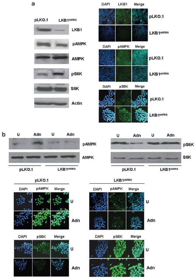
Adiponectin is widely known as an adipocytokine with therapeutic potential for its markedly protective function in the pathogenesis of obesity-related disorders, metabolic syndrome, systemic insulin resistance, cardiovascular disease and more recently carcinogenesis. In the present study, we show that adiponectin inhibits adhesion, invasion and migration of breast cancer cells. Further analysis of the underlying molecular mechanisms revealed that adiponectin treatment increased AMP-activated protein kinase (AMPK) phosphorylation and activity as evident by increased phosphorylation of downstream target of AMPK, acetyl-coenzyme A carboxylase and inhibition of p70S6 kinase (S6K). Intriguingly, we discovered that adiponectin treatment increases the expression of tumor suppressor gene LKB1 in breast cancer cells. Overexpression of LKB1 in breast cancer cells further increased adiponectin-mediated phosphorylation of AMPK. Using isogenic LKB1 knockdown cell line pair, we found that LKB1 is required for adiponectin-mediated modulation of AMPK-S6K axis and more importantly, inhibition of adhesion, migration and invasion of breast cancer cells. Taken together these data present a novel mechanism involving specific upregulation of tumor suppressor gene LKB1 by which adiponectin inhibits adhesion, invasion and migration of breast cancer cells. Our findings indicate the possibility of using adiponectin analogues to inhibit invasion and migration of breast cancer cells.


Conflict of interest statement

Conflict of interest

The authors declare no conflict of interest.

Figures

Figure 1
Figure 1
Adiponectin inhibits adhesion, migration and invasion of breast cancer cells. (a)MCF7 and T47D breast cancer cells were grown to confluence, scratched with a pipette tip and were photographed immediately following scratching (0 h). Culture media were replaced with media containing 10 μg/ml adiponectin (Adn)or untreated media (U). The plates were photographed at the identical location of the initial image (0 h)at 24 and 48 h. The results shown are representative of three independent experiments performed in triplicates. Adiponectin inhibited migration of breast cancer cells. (b)Confluent layer of MCF7 and T47D breast cancer cells grown on electric cell-substrate impedance sensing (ECIS) 8W1E plates was subjected to an elevated voltage pulse of 40 kHz frequency, 3.5V amplitude for 30 s duration to create wound and resistance was measured for 24 h in the presence (Adn)and absence (untreated)of 10 μg/ml adiponectin to follow migration of breast cancer cells. Adiponectin treatment inhibited migration of breast cancer cells in an ECIS assay. All the experiments were performed thrice in triplicates. (c)MCF7 and T47D cells were plated at a density of 250 cells per well in 12-well plates and grown in the presence (Adn)or absence (U) of 10 μg/ml of adiponectin for 1 week. Cells were fixed and stained with crystal violet. The results shown are representative of three independent experiments performed in triplicates. Colonies containing >50 normal-appearing cells were counted. The histogram is the number of colonies. *P<0.001, compared with untreated cells. Adiponectin treatment significantly reduced colony formation by MCF7 and T47D breast cancer cells. (d)MCF7 and T47D cells were pretreated with 10 μg/ml of adiponectin for 24 h and plated in 10 μg/cm2 fibronectin-coated wells in a 96-well plate for 60 min. The adherent cells were stained with crystal violet. The stain was eluted and absorbance was measured at 540 nm. The data represent mean values±s.e.m. and are the results of three independent experiments. *P<0.005, compared with untreated control cells. Adiponectin treatment significantly reduced adhesion. (e)MCF7 and T47D cells were cultured in Matrigel invasion chambers followed by treatment with 10 μg/ml of adiponectin for 24 h. The number of cells that invaded through the Matrigel was counted in five different regions. The slides were blinded to remove counting bias. The result shows mean of three independent experiments performed in triplicates. *P<0.005, compared with untreated controls. Adiponectin treatment significantly reduced Matrigel invasion.
Figure 2
Figure 2
Evidence of adiponectin-induced activation of AMP-activated protein kinase (AMPK). (a)MCF7 and T47D cells were treated with 10 μg/ml of adiponectin for indicated time intervals. Untreated cells are denoted with 0. Total protein was isolated and equal amounts of proteins were resolved by SDS–polyacrylamide gel (PAGE)and subjected to immunoblot analysis using specific antibodies for phosphorylated AMPK (pAMPK-Thr172). The membranes were reblotted using total AMPK antibody as control. The blots are representative of multiple independent experiments. The histogram is the mean of densitometric analysis showing relative density units (RDU)of the western blot signal for pAMPK normalized to total AMPK in three separate experiments. *P<0.005, compared with untreated controls. Adiponectin treatment increases phosphorylation of AMPK in MCF7 and T47D breast cancer cells. (b)MCF7 and T47D cells were treated with 10 μg/ml of adiponectin (Adn)for 2 h and phosphorylation of AMPK was analysed by immunofluorescence using pAMPK-Thr172 antibody. 4′,6-Diamidino-2-phenylindole (DAPI)staining was used to determine the nuclear localization. These results are representative of multiple independent experiments. Adiponectin treatment increased nuclear accumulation of phosphorylated AMPK. (c)MCF7 and T47D cells were treated with 10 μg/ml of adiponectin for indicated time intervals. Untreated cells are denoted with 0. Total protein was isolated and equal amounts of proteins were resolved by SDS–PAGE and subjected to immunoblot analysis using specific antibodies for phosphorylated ACC (pACC). The membranes were reblotted using total ACC antibody as control. The blots are representative of multiple independent experiments. The histogram is the mean of densitometric analysis showing RDU of the western blot signals for pACC normalized to total ACC in three independent experiments. *P<0.005, compared with untreated controls. Adiponectin treatment increases phosphorylation of ACC in MCF7 and T47D breast cancer cells. (d)MCF7 and T47D cells were treated with 10 μg/ml of adiponectin (Adn)for 2 h and phosphorylation of ACC was analysed by immunofluorescence using pACC antibody. DAPI staining was used to determine the nuclear localization. These results are representative of multiple independent experiments. Adiponectin treatment increased nuclear accumulation of pACC.
Figure 3
Figure 3
Adiponectin decreases phosphorylation of S6K in breast cancer cells. (a)MCF7 and T47D cells were treated with 10 μg/ml of adiponectin for indicated time intervals. Untreated cells are denoted with 0. Total protein was isolated and equal amounts of proteins were resolved by SDS–polyacrylamide gel (PAGE)and subjected to immunoblot analysis using specific antibodies for phosphorylated S6K (pS6K). The membranes were reblotted using total S6K antibody as control. The blots are representative of multiple independent experiments. The histogram is the mean of densitometric analysis showing relative density units (RDU)of the western blot signals for pS6K normalized to total S6K in three separate experiments. *P<0.001, compared with untreated controls. Adiponectin treatment decreases phosphorylation of S6K in MCF7 and T47D breast cancer cells. (b)MCF7 and T47D cells were treated with 10 μg/ml of adiponectin (Adn)for 4 h and phosphorylation of S6K was analysed by immunofluorescence using pS6K antibody. 4′,6-Diamidino-2-phenylindole (DAPI)staining was used to determine the nuclear localization. These results are representative of multiple independent experiments. Adiponectin treatment decreased overall (nuclear and cytoplasmic)accumulation of pS6K.
Figure 4
Figure 4
Adiponectin function involves upregulation of LKB1. (a)MCF7 and T47D cells were treated with 10 μg/ml of adiponectin for indicated time intervals. Untreated cells are denoted with 0. Total protein was isolated and equal amounts of proteins were resolved by SDS–polyacrylamide gel (PAGE)and subjected to immunoblot analysis using specific antibodies for LKB1. The membranes were reblotted using actin antibody as control. The blots are representative of multiple independent experiments. The histogram is the mean of densitometric analysis showing relative density units (RDU)of the western blot signals for LKB1 normalized to actin in three independent experiments. *P<0.005, compared with untreated controls. Adiponectin treatment increases level of LKB1 protein in MCF7 and T47D breast cancer cells. (b)MCF7 and T47D cells were treated with 10 μg/ml of adiponectin (Adn)for 2 h and LKB1 protein was analysed by immunofluorescence using LKB1 antibody. 4′,6-Diamidino-2-phenylindole (DAPI)staining was used to determine the nuclear localization. These results are representative of multiple independent experiments. Adiponectin treatment increased cytoplasmic LKB1 levels in breast cancer cells. (c)MCF7 and T47D cells were transfected with Flag-tagged wild-type LKB1 (LKB1)or vector control (C)using Lipofectamine 2000 (Invitrogen)for 48 h. Total protein was isolated and equal amounts of proteins were resolved by SDS–PAGE and subjected to immunoblot analysis using specific antibodies for LKB1, Flag, pAMPK-Thr172, AMPK, pS6K, S6K and actin. These results are representative of multiple independent experiments. Overexpression of LKB1 increases phosphorylation of AMPK and reduces phosphorylation of S6K. (d)MCF7 cells were transfected with Flag-tagged wild-type LKB1 (WT-LKB1)or vector control (Ctrl) using Lipofectamine 2000 (Invitrogen) for 40 h followed by treatment with 10 μg/ml of adiponectin (Adn)for indicated time intervals. Total protein was isolated and equal amounts of proteins were resolved by SDS–PAGE and subjected to immunoblot analysis using specific antibodies for phosphorylated AMPK (pAMPK-Thr172). The membranes were reblotted using total AMPK antibody as control. The blots are representative of multiple independent experiments. The histogram is the mean of densitometric analysis showing RDU of the western blot signals (pAMPK) normalized to total AMPK in three separate experiments. *P<0.005, compared with untreated controls. #P<0.001 compared with untreated controls and **P<0.005, compared to untreated controls. Overexpression of LKB1 further increases adiponectin-induced phosphorylation of AMPK.
Figure 5
Figure 5
Depletion of LKB1 in MCF7 cells abrogates adiponectin-mediated modulation of AMPK–S6K axis. (a)LKB1 was depleted in MCF7 cells using lentiviral LKB1 short-hairpin RNA (shRNA)constructs and a negative control construct that was created in the same vector system (pLKO.1)(Open Biosystems). Stable pools of LKB1-depleted (LKB1shRNA)and vector control (pLKO.1)cells were used for total protein isolation and equal amounts of proteins were subjected to immunoblot analysis using specific antibodies for LKB1, phosphorylated AMPK (pAMPK), AMP-activated protein kinase (AMPK), phosphorylated S6K (pS6K), S6K and actin. The blots are representative of multiple independent experiments. LKB1shRNA and pLKO.1 cells were analysed by immunofluorescence using LKB1, pAMPK and pS6K antibodies. 4′,6-Diamidino-2-phenylindole (DAPI)staining was used to determine the nuclear localization. Depletion of LKB1 inhibits nuclear accumulation of pAMPK and increases nuclear and cytoplasmic accumulation of pS6K. (b)LKB1 shRNA and pLKO.1 cells were treated with 10 μg/ml of adiponectin (Adn)for 2 h and phosphorylation of AMPK and S6K was analysed by western blot analysis and immunofluorescence using pAMPK-Thr172 and pS6K antibody. DAPI staining was used to determine the nuclear localization. These results are representative of multiple independent experiments. Adiponectin treatment increased phosphorylation of AMPK and decreased phosphorylation of S6K in pLKO.1 cells whereas no effect of adiponectin was observed in LKB1shRNA cells.
Figure 6
Figure 6
Depletion of LKB1 in MCF7 cells abrogates adiponectin-mediated inhibition of adhesion, invasion and migration of breast cancer cells. (a)LKB1 shRNA and pLKO.1 cells were grown to confluence, scratched with a pipette tip and were photographed immediately following scratching (0 h). Culture media were replaced with media containing 10 μg/ml adiponectin (Adn)or untreated media (U). The plates were photographed at the identical location of the initial image (0 h) at 24 and 48 h. The results shown are representative of three independent experiments performed in triplicates. Adiponectin inhibited migration of pLKO.1 cells whereas LKB1shRNA cells remain unaffected. (b)Confluent layer of LKB1shRNA and pLKO.1 cells grown on electric cell-substrate impedance sensing (ECIS) 8W1E plates were subjected to an elevated voltage pulse of 40 kHz frequency, 3.5V amplitude for 30 s duration to create wound and resistance was measured for 24 h in the presence (Adn)and absence (untreated)of 10 μg/ml adiponectin to follow migration of LKB1shRNA and pLKO.1 cells. Adiponectin treatment inhibited migration of pLKO.1 cells whereas LKB1shRNA cells show no effect of adiponectin in an ECIS assay. All the experiments were performed thrice in triplicates. (c)LKB1 shRNA and pLKO.1 cells were plated at a density of 250 cells per well in 12-well plates and grown in the presence (Adn)or absence (U)of 10 μg/ml of adiponectin for 1 week. Cells were fixed and stained with crystal violet. The results shown are representative of three independent experiments performed in triplicates. Colonies containing >50 normal-appearing cells were counted. The histogram is the number of colonies. *P<0.001, compared with untreated cells. Adiponectin treatment significantly reduced colony formation by pLKO.1 cells whereas LKB1shRNA cells remain largely unaffected. (d)LKB1 shRNA and pLKO.1 cells were pretreated with 10 μg/ml of adiponectin for 24 h and plated in 10 μg/cm2 fibronectin-coated wells in a 96-well plate for 60 min. The adherent cells were stained with crystal violet. The stain was eluted and absorbance was measured at 540 nm. The data represent mean values±s.e.m. and are the results of three independent experiments. *P<0.005, compared with untreated control cells. Adiponectin treatment significantly reduced adhesion of pLKO.1 cells whereas LKB1shRNA cells remain unaffected. (e)LKB1 shRNA and pLKO.1 cells were cultured in Matrigel invasion chambers followed by treatment with 10 μg/ml of adiponectin for 24 h. The number of cells that invaded through the Matrigel was counted in five different regions. The slides were blinded to remove counting bias. The result shows mean of three independent experiments performed in triplicates. *P<0.005, compared with untreated controls. Adiponectin treatment significantly reduced invasion of pLKO.1 cells while LKB1shRNA cells remain mostly unaffected.

Similar articles

Cited by

References

    1. Alessi DR, Sakamoto K, Bayascas JR. LKB1-dependent signaling pathways. Annu Rev Biochem. 2006;75:137–163. - PubMed
    1. Alimova IN, Liu B, Fan Z, Edgerton SM, Dillon T, Lind SE, et al. Metformin inhibits breast cancer cell growth, colony formation and induces cell cycle arrest in vitro. Cell Cycle. 2009;8:909–915. - PubMed
    1. Arditi JD, Venihaki M, Karalis KP, Chrousos GP. Antiproliferative effect of adiponectin on MCF7 breast cancer cells: a potential hormonal link between obesity and cancer. Horm Metab Res. 2007;39:9–13. - PubMed
    1. Baas AF, Boudeau J, Sapkota GP, Smit L, Medema R, Morrice NA, et al. Activation of the tumour suppressor kinase LKB1 by the STE20-like pseudokinase STRAD. EMBO J. 2003;22:3062–3072. - PMC - PubMed
    1. Barb D, Pazaitou-Panayiotou K, Mantzoros CS. Adiponectin: a link between obesity and cancer. Expert Opin Investig Drugs. 2006;15:917–931. - PubMed

Publication types

MeSH terms