. 2009 Jul 16;28(28):2581-92.
doi: 10.1038/onc.2009.124. Epub 2009 Jun 1.

Pim-1 plays a pivotal role in hypoxia-induced chemoresistance

Affiliations

Pim-1 plays a pivotal role in hypoxia-induced chemoresistance

J Chen et al. Oncogene. .

Abstract

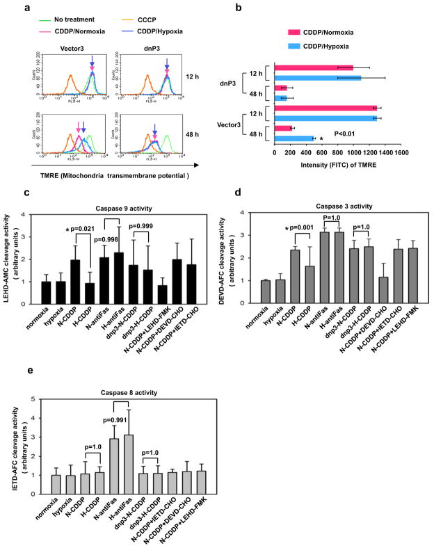
Hypoxia changes the responses of cancer cells to many chemotherapy agents, resulting in chemoresistance. The underlying molecular mechanism of hypoxia-induced drug resistance remains unclear. Pim-1 is a survival kinase, which phosphorylates Bad at serine 112 to antagonize drug-induced apoptosis. Here we show that hypoxia increases Pim-1 in a hypoxia-inducible factor-1alpha-independent manner. Inhibition of Pim-1 function by dominant-negative Pim-1 dramatically restores the drug sensitivity to apoptosis induced by chemotherapy under hypoxic conditions in both in vitro and in vivo tumor models. Introduction of siRNAs for Pim-1 also resensitizes cancer cells to chemotherapy drugs under hypoxic conditions, whereas forced overexpression of Pim-1 endows solid tumor cells with resistance to cisplatin, even under normoxia. Dominant-negative Pim-1 prevents a decrease in mitochondrial transmembrane potential in solid tumor cells, which is normally induced by cisplatin (CDDP), followed by the reduced activity of Caspase-3 and Caspase-9, indicating that Pim-1 participates in hypoxia-induced drug resistance through the stabilization of mitochondrial transmembrane potential. Our results demonstrate that Pim-1 is a pivotal regulator involved in hypoxia-induced chemoresistance. Targeting Pim-1 may improve the chemotherapeutic strategy for solid tumors.


Conflict of interest statement

Conflict of interest

The authors declare no conflict of interest.

Figures

Figure 1
Figure 1. Hypoxia induces resistance to several cytotoxic drugs in tumor cells
Representative results from three independent experiments are shown. (a) Gemcitabine dose-response curves under normoxic or hypoxic conditions for PCI-43 cells treated with indicated concentrations of gemcitabine for 48 hours. Cell viability was estimated by colorimetric MTS assay. Error bars represent standard deviation. (b) Doxorubicin dose-response curves in PCI-43 cells under normoxic or hypoxic conditions for PCI-43 cells treated with indicated concentrations of doxorubicin for 48 hours. (c) PCI-43 cells treated with CDDP (25 μM) were cultured under hypoxic conditions for the indicated times and percentage of apoptotic cells determined by PI and annexin V staining using FACS analysis. (d) PCI-43 cells treated with CDDP (25 μM) for 72 hours under hypoxic or normoxia conditions, cell cycles were measured by PI staining using FACS analysis. (e) KMP-4, HeLa and PCI-35 cells were treated with CDDP (25 μM) for 48 hours and the percentage of apoptotic cells determined in the same manner as in (c). (f) PCI-43 cells were treated with fluorouracil (100 μM) and A23187 (5 μM) for 48 hours. Apoptosis and cell viability were assessed using FACS as described in (c). (g) HeLa cells were treated with indicated chemotherapeutic drugs for 48 h under normoxic or hypoxic conditions and dead cell fraction determined as described in (c).
Figure 2
Figure 2. Pim-1 is up-regulated by hypoxia in a HIF-1α-independent manner
(a) A panel of cell lines was cultured under hypoxic or normoxic conditions for 16 hours. Western and Northern blot analyses for Pim-1 are shown. (b) Western blot analyses of Bcl-2 family proteins and Pim-2 in PCI-43 cells following incubation under hypoxia at the indicated times. (c) Western blot analyses of HIF-1α, VEGF, Glut-1 and Pim-1 following incubation under hypoxia at the indicated times in PCI-43 cells (left), and in dominant negative HIF-1α (dnHIF-1α) transfectant or vector control (vector3) (right). (d) Pim-1 induced by hypoxia in HIF-1α knock down cells. HeLa cells or HCT116 cells were transfected with HIF-1α siRNAs for 72 hours, and then were incubated under normoxia or hypoxia for 6 hours. The Pim-1 protein and mRNA expression were analyzed by Western blot assay and quantitative PCR, respectively. β-actin mRNA was used to standardize the total amount of cDNA. * indicates significance (P<0.01). (e) 32P-labeled double stranded oligonucleotide probes (EPO wt33, wild type probes of proPim-1HRE1, proPim-1HRE2, proPim-1HRE3 and proPim-1HRE4) containing the consensus HIF-1 binding site were incubated with nuclear extracts from PCI-43 cells cultured under normoxia or hypoxia for 16 h and analyzed by EMSA. For the competition assay (comp), the wild type (W) or mutant (M) unlabeled oligonucleotide probes were added to nuclear extracts before the addition of labeled probe in normoxia. N: normoxia, H: hypoxia. (f) Luciferase reporter assay for co-transfectants of pGL3-proPim-1 (−1715 to +38bp) with HIF-1α in normoxia (black bar, line2). pGL3-5×HRE and HIF-1α were co-transfected in normoxia as a positive control. pGL3-proPim-1 or pGL3-5×HRE was transfected in hypoxia as another control. 293 cells were co-transfected with indicated plasmids for 24 h. After incubation in normoxia or hypoxia for 16 h, luciferase activity was measured. Fold induction relative to Propim-1 is plotted. Results shown are from three independent experiments.
Figure 3
Figure 3. Dominant negative Pim-1 (dnPim-1) sensitizes cells to apoptosis induced by CDDP in the presence of hypoxia
Representative results of FACS analysis for three independent experiments are shown. (a) Structures of wild type Pim-1 and dominant negative Pim-1 (dnPim-1). (b) Expression of various dnPim-1 proteins in PCI-43 transfectants. Anti-HA immunoblot is shown. * indicates a non-specific band. (c) Western blot of Bad and phosphorylated Bad-ser112 protein following incubation under hypoxia for 12 hours in dnPim-1 transfectants. (d) Apoptotic analysis of dnPim-1-transfectants (dnp3, 4) without drug treatment for 48 hours in the presence of normoxia or hypoxia. Apoptotic cells were quantified by PI and annexin V staining via FACS analysis. (e) Apoptotic analysis of the dnPim-1-transfectants treated with CDDP (25 μM) for 48 hours in the presence of normoxia or hypoxia. Apoptotic cells were quantified as described in (d). (f) Apoptotic analysis of the dnPim-1-transfectants treated with anti-Fas Ab for 48 hours in the presence of normoxia or hypoxia. Apoptotic cells were quantified as described in (d).
Figure 4
Figure 4. Pim-1 siRNA or Pim-1 overexpression has impact on apoptotic response
Representative results of FACS analysis for three independent experiments are shown. (a) Expression of Pim-1 proteins in PCI-43 cells treated with Pim-1 siRNAs under normoxia and hypoxia (12 hours). N: normoxia, H: hypoxia. (b) Apoptotic analysis of cells treated with Pim-1 siRNAs, followed by CDDP (25 μM) for 48 hours in the presence of normoxia and hypoxia. Apoptotic cells were quantified by PI and annexin V staining via FACS analysis. (c) Apoptotic cell percentage in three independent experiments of b. The error bars represent SD. * indicates significance (P<0.01). (d) Western blot of Pim-1 proteins in PCI-43 Pim-1 transfectants. Anti-Flag immunoblot is shown. * indicates a non-specific band. (e) Apoptotic analysis of Pim-1 transfectants treated with CDDP (25 μM) for 48 hours under normoxia. Apoptotic cells were quantified as described in (b). (f) Apoptotic cell percentage in three independent experiments of e. The error bars represent SD. * indicates significance (P<0.01).
Figure 5
Figure 5. In vivo sensitivity of the Tet-on dnPim-1-transfectant to CDDP
(a) Western blot analyses of dnPim-1 in Tet-on dnPim-1-transfectants. (b) Growth of Tet-On dnPim-1 transfectants in SCID mice treated with CDDP. Five mice in each group were inoculated with 5×10⁶ tumor cells on Day 0. Doxycycline administration was initiated on Day 4 by adding to drinking water at 2 μg/ml delivering approximately 13 mg/kg/day. CDDP (2 mg/kg) was injected intra-peritoneally once per week beginning on Day 4. Tumor size was measured every three days following inoculation. The error bars represent SD, * indicates significance (P<0.05) compared to dnPim-1/DOX(−) control.
Figure 6
Figure 6. Pim-1 induces resistance to anti-cancer drugs through stabilization of mitochondrial transmembrane potential
Representative results for three independent experiments are shown. (a) Mitochondrial transmembrane potential (MTP) examined in dnPim-1-transfectants treated with 25 μM CDDP under normoxia and hypoxia. TMRE (200 nM) was added to indicate MTP during the last 30 minutes of treatment with hypoxia or normoxia, followed by FACS analysis. Untreated cells (negative control) and cells treated with 50 μM CCCP (inhibitor of MTP, positive control) were included in the assays. (b) Quantification of mitochondrial transmembrane potential (MTP) in (a). * indicates significance (P<0.01). (c) dnPim-1 transfectants under normoxic or hypoxic conditions with the indicated treatments were assayed for Caspase-9 activity via spectrofluorometer. (d) Caspase-3 activity was assayed as described in (c). (e) Caspase-8 activity was assayed as described in (c). The activities of Caspase-3,-8, and-9 were inhibited with Ac-DEVD-CHO, Ac-IETD-CHO and Z-LEHD-FMK respectively. N: normoxia, H: hypoxia.

References

    1. Aho TL, Sandholm J, Peltola KJ, Mankonen HP, Lilly M, Koskinen PJ. Pim-1 kinase promotes inactivation of the pro-apoptotic Bad protein by phosphorylating it on the Ser112 gatekeeper site. FEBS Lett. 2004;571:43–9. - PubMed
    1. Arsham AM, Plas DR, Thompson CB, Simon MC. Akt and hypoxia-inducible factor-1 independently enhance tumor growth and angiogenesis. Cancer Res. 2004;64:3500–7. - PubMed
    1. Bandyopadhyay RS, Phelan M, Faller DV. Hypoxia induces AP-1-regulated genes and AP-1 transcription factor binding in human endothelial and other cell types. Biochim Biophys Acta. 1995;1264:72–8. - PubMed
    1. Cardenas-Navia LI, Yu D, Braun RD, Brizel DM, Secomb TW, Dewhirst MW. Tumor-dependent kinetics of partial pressure of oxygen fluctuations during air and oxygen breathing. Cancer Res. 2004;64:6010–7. - PubMed
    1. Chen J, Kobayashi M, Darmanin S, Qiao Y, Gully C, Zhao R, et al. Hypoxia-mediated upregulation of Pim-1 contributes to solid tumor formation. Am J Pathol. 2009 in press. - PMC - PubMed

Publication types

MeSH terms

Substances