Review
. 2009 Oct;66(20):3241-61.
doi: 10.1007/s00018-009-0051-1. Epub 2009 May 31.

Mammalian iron transport

Affiliations
Review

Mammalian iron transport

Gregory Jon Anderson et al. Cell Mol Life Sci. 2009 Oct.

Abstract

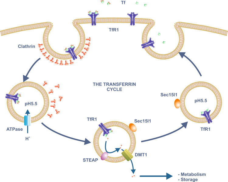
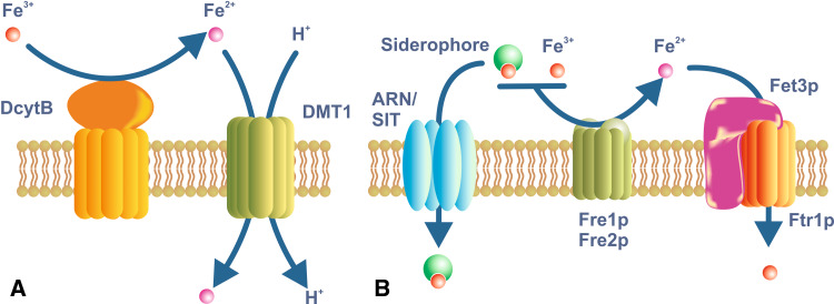
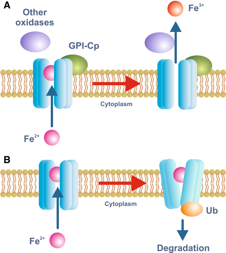
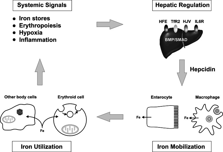
Iron is essential for basic cellular processes but is toxic when present in excess. Consequently, iron transport into and out of cells is tightly regulated. Most iron is delivered to cells bound to plasma transferrin via a process that involves transferrin receptor 1, divalent metal-ion transporter 1 and several other proteins. Non-transferrin-bound iron can also be taken up efficiently by cells, although the mechanism is poorly understood. Cells can divest themselves of iron via the iron export protein ferroportin in conjunction with an iron oxidase. The linking of an oxidoreductase to a membrane permease is a common theme in membrane iron transport. At the systemic level, iron transport is regulated by the liver-derived peptide hepcidin which acts on ferroportin to control iron release to the plasma.


Figures

Fig. 1
Fig. 1
The transferrin cycle. Plasma ferric iron binds with high affinity to Tf which in turn binds to TfR1 on the plasma membrane. The Tf/TfR1 complex is internalized through clathrin-coated pits by receptor-mediated endocytosis. A proton-pumping ATPase lowers the pH of the endosome to around 5.5. Iron is released at this low pH, a process aided by reduction of the ferric iron by enzymes of the STEAP family. The resulting ferrous iron moves across the endosomal membrane via DMT1 and enters the cytoplasm where it can be utilized for various metabolic functions or stored within ferritin. ApoTf remains bound to TfR1 at the low pH of the endosome and is returned to the extracellular medium after the endosome recycles to the plasma membrane. The trafficking protein Sec15l1 is involved in Tf recycling. Abbreviations: see text
Fig. 2
Fig. 2
Iron uptake in enterocytes and yeast. a Dietary iron is taken up across the brush border membrane of duodenal enterocytes through the iron/proton symporter DMT1. DMT1 is expressed on most body cells, but it plays a specialized role in iron acquisition from the diet. Most dietary iron is in the Fe 3+ or ferric form, but it needs to be reduced to the Fe 2+ or ferrous form before it can be utilized as a substrate by DMT1. DcytB is an iron-regulated reductase of the brush border membrane that is a candidate for this role. However, whether DcytB is the only reductase involved remains to be determined. b The yeast Saccharomyces cerevisiae is a simple eukaryote that is able to utilize iron in a variety of forms. It also must reduce iron before it can be utilized. The reductases Fre1p and Fre2p are the principal plasma membrane enzymes involved in this process, but several others have been described. They can utilize both ionic iron or siderophore-bound iron as substrates. The reduced iron is transported into the cell through a complex of the iron permease Ftr1p and the iron oxidase Fet3p. A variety of siderophores also can be utilized by S. cerevisiae, and these are taken up via members of the ARN and SIT families
Fig. 3
Fig. 3
Model for iron export through FPN. Iron export through FPN requires the action of an iron oxidase and this function is usually fulfilled by either circulating or GPI-linked forms of Cp. De Domenico et al. [130] have proposed a model for the function of Cp whereby the oxidation of FPN-bound ferrous iron by Cp is required for its release into the extracellular medium (a). In the absence of appropriate oxidase activity, ferrous iron remains bound to FPN and the protein is ubiquitinated and targeted for degradation (b). This is only one mechanism by which FPN expression on the plasma membrane can be modulated. The binding of hepcidin to FPN can also facilitate its internalization and degradation
Fig. 4
Fig. 4
The regulation of body iron metabolism. The amount of iron entering the plasma is controlled by body iron demand and this in turn reflects a range of stimuli. Low body iron levels, increased erythropoiesis and hypoxia are all stimuli for increased iron export into the circulation from the tissues. Inflammation has the opposite effect and leads to tissue iron withholding. These stimuli exert their effects by acting through a series of proteins on the hepatocyte plasma membrane, including HFE, TfR2, HJV and the IL-6 receptor, to modulate the expression of the iron regulatory hormone hepcidin. Hepcidin in turn binds to FPN on the surface of target cells to influence how much iron they release. Enterocytes and macrophages are major targets, but it is likely that most body cells can respond to hepcidin. An increased hepcidin level leads to reduced FPN on the target membrane and reduced iron release. Iron that enters the plasma is used by all body cells for metabolic functions, but immature erythroid cells have particularly high iron requirements for hemoglobin synthesis. It is the iron requirements of these target cells which complete the cycle

Similar articles

Cited by

References

    1. Crosa JH, Mey AR, Payne SM, editors. Iron transport in bacteria. Washington, DC: American Society for Microbiology; 2004.
    1. Philpott CC, Protchenko O. Response to iron deprivation in Saccharomyces cerevisiae . Eukaryot Cell. 2008;7:20–27. doi: 10.1128/EC.00354-07. - DOI - PMC - PubMed
    1. Sutak R, Lesuisse E, Tachezy J, Richardson DR. Crusade for iron: iron uptake in unicellular eukaryotes and its significance for virulence. Trends Microbiol. 2008;16:261–268. doi: 10.1016/j.tim.2008.03.005. - DOI - PubMed
    1. Wilson MT, Reeder BJ. Oxygen-binding haem proteins. Exp Physiol. 2008;93:128–132. doi: 10.1113/expphysiol.2007.039735. - DOI - PubMed
    1. Rouault TA, Tong WH. Iron-sulphur cluster biogenesis and mitochondrial iron homeostasis. Nat Rev Mol Cell Biol. 2005;6:345–351. doi: 10.1038/nrm1620. - DOI - PubMed

MeSH terms