Correlation between gross anatomical topography, sectional sheet plastination, microscopic anatomy and endoanal sonography of the anal sphincter complex in human males
- PMID: 19486204
- PMCID: PMC2740969
- DOI: 10.1111/j.1469-7580.2009.01091.x
Correlation between gross anatomical topography, sectional sheet plastination, microscopic anatomy and endoanal sonography of the anal sphincter complex in human males
Abstract
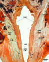
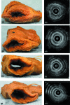
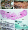
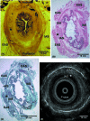
This study elucidates the structure of the anal sphincter complex (ASC) and correlates the individual layers, namely the external anal sphincter (EAS), conjoint longitudinal muscle (CLM) and internal anal sphincter (IAS), with their ultrasonographic images. Eighteen male cadavers, with an average age of 72 years (range 62-82 years), were used in this study. Multiple methods were used including gross dissection, coronal and axial sheet plastination, different histological staining techniques and endoanal sonography. The EAS was a continuous layer but with different relations, an upper part (corresponding to the deep and superficial parts in the traditional description) and a lower (subcutaneous) part that was located distal to the IAS, and was the only muscle encircling the anal orifice below the IAS. The CLM was a fibro-fatty-muscular layer occupying the intersphincteric space and was continuous superiorly with the longitudinal muscle layer of the rectum. In its middle and lower parts it consisted of collagen and elastic fibres with fatty tissue filling the spaces between the fibrous septa. The IAS was a markedly thickened extension of the terminal circular smooth muscle layer of the rectum and it terminated proximal to the lower part of the EAS. On endoanal sonography, the EAS appeared as an irregular hyperechoic band; CLM was poorly represented by a thin irregular hyperechoic line and IAS was represented by a hypoechoic band. Data on the measurements of the thickness of the ASC layers are presented and vary between dissection and sonographic imaging. The layers of the ASC were precisely identified in situ, in sections, in isolated dissected specimens and the same structures were correlated with their sonographic appearance. The results of the measurements of ASC components in this study on male cadavers were variable, suggesting that these should be used with caution in diagnostic and management settings.
Figures


Similar articles
-
Histo-topographic study of the longitudinal anal muscle.Clin Anat. 2008 Jul;21(5):447-52. doi: 10.1002/ca.20633. Clin Anat. 2008. PMID: 18561297
-
The role of the longitudinal muscle in the anal sphincter complex: Implications for the Intersphincteric Plane in Low Rectal Cancer Surgery?Clin Anat. 2020 May;33(4):567-577. doi: 10.1002/ca.23444. Epub 2019 Aug 30. Clin Anat. 2020. PMID: 31385374
-
Correlation of endoanal sonography with cross-sectional anatomy of the anal sphincter.Gastrointest Endosc. 1999 Dec;50(6):804-10. doi: 10.1016/s0016-5107(99)70162-8. Gastrointest Endosc. 1999. PMID: 10570340
-
Anal canal anatomy showed by three-dimensional anorectal ultrasonography.Surg Endosc. 2007 Dec;21(12):2207-11. doi: 10.1007/s00464-007-9339-0. Epub 2007 May 4. Surg Endosc. 2007. PMID: 17479327
-
The anatomical basis of anal endosonography. A study in postmortem specimens.Surg Endosc. 1997 Oct;11(10):986-90. doi: 10.1007/s004649900508. Surg Endosc. 1997. PMID: 9381354
Cited by
-
The Female Pelvic Floor Fascia Anatomy: A Systematic Search and Review.Life (Basel). 2021 Aug 30;11(9):900. doi: 10.3390/life11090900. Life (Basel). 2021. PMID: 34575049 Free PMC article. Review.
-
Morphology of the region anterior to the anal canal in males: visualization of the anterior bundle of the longitudinal muscle by transanal ultrasonography.Surg Radiol Anat. 2017 Sep;39(9):967-973. doi: 10.1007/s00276-017-1832-0. Epub 2017 Feb 28. Surg Radiol Anat. 2017. PMID: 28247085
-
Generation of myogenic progenitor cell-derived smooth muscle cells for sphincter regeneration.Stem Cell Res Ther. 2020 Jun 12;11(1):233. doi: 10.1186/s13287-020-01749-w. Stem Cell Res Ther. 2020. PMID: 32532320 Free PMC article.
-
A New Approach to Ultra-Low Anterior Resection-Intersphincteric Dissection With Total Hiatal Ligament Excision for Very Low Rectal Cancer Located in the Posterior Wall of the Rectum: A More Satisfactory Technique for Local Recurrence Control.Cancer Med. 2024 Oct;13(19):e70307. doi: 10.1002/cam4.70307. Cancer Med. 2024. PMID: 39387535 Free PMC article.
References
-
- Ayoub SF. Anatomy of the external anal sphincter in man. Acta Anat (Basel) 1979;105:25–36. - PubMed
-
- Beets-Tan RG, Morren GL, Beets GL, et al. Measurement of anal sphincter muscles: endoanal US, endoanal MR imaging, or phased-array MR imaging? A study with healthy volunteers. Radiology. 2001;220:81–89. - PubMed
-
- Cook P, Al-Ali SYA. Submacroscopic interpretation of human sectional anatomy using plastinated E12 sections. J Int Soc Plast. 1997;12:17–27.
-
- Cornella JL, Hibner M, Fenner DE, Kriegshauser JS, Hentz J, Magrina JF. Three-dimensional reconstruction of magnetic resonance images of the anal sphincter and correlation between sphincter volume and pressure. Am J Obstet Gynecol. 2003;189:130–135. - PubMed
-
- Courtney H. Anatomy of the pelvic diaphragm and anorectal musculature as related to sphincter preservation in anorectal surgery. Am J Surg. 1950;79:155–173. - PubMed
Publication types
MeSH terms
Substances
LinkOut - more resources
Full Text Sources
Other Literature Sources
Miscellaneous

