. 2010 Jan;48(1):238-45.
doi: 10.1016/j.yjmcc.2009.05.014. Epub 2009 May 30.

Connexin40 and connexin43 determine gating properties of atrial gap junction channels

Affiliations

Connexin40 and connexin43 determine gating properties of atrial gap junction channels

Xianming Lin et al. J Mol Cell Cardiol. 2010 Jan.

Abstract

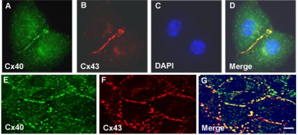
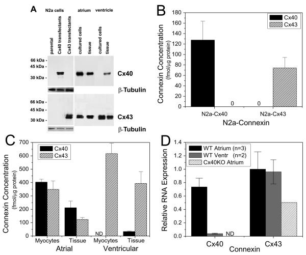
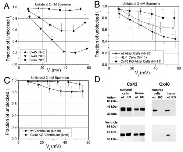
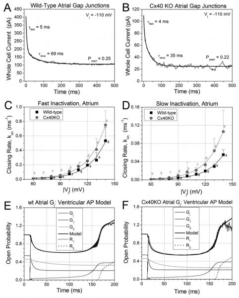
While ventricular gap junctions contain only Cx43, atrial gap junctions contain both Cx40 and Cx43; yet the functional consequences of this co-expression remain poorly understood. We quantitated the expression of Cx40 and Cx43 and their contributions to atrial gap junctional conductance (g(j)). Neonatal murine atrial myocytes showed similar abundances of Cx40 and Cx43 proteins, while ventricular myocytes contained at least 20 times more Cx43 than Cx40. Since Cx40 gap junction channels are blocked by 2 mM spermine while Cx43 channels are unaffected, we used spermine block as a functional dual whole cell patch clamp assay to determine Cx40 contributions to cardiac g(j). Slightly more than half of atrial g(j) and <or=20% of ventricular g(j) were inhibited. In myocytes from Cx40 null mice, the inhibition of ventricular g(j) was completely abolished, and the block of atrial g(j) was reduced to <20%. Compared to ventricular gap junctions, the transjunctional voltage (V(j))-dependent inactivation of atrial g(j) was reduced and kinetically slowed, while the V(j)-dependence of fast and slow inactivation was unchanged. We conclude that Cx40 and Cx43 are equally abundant in atrium and make similar contributions to atrial g(j). Co-expression of Cx40 accounts for most, but not all, of the differences in the V(j)-dependent gating properties between atrium and ventricle that may play a role in the genesis of slow myocardial conduction and arrhythmias.


Figures

Figure 1
Figure 1
Immunofluorescent localization of Cx40 and Cx43 in neonatal mouse atrial cultures. A-D, atrial myocyte cell pair showing immunolocalization of Cx40 (green in A), Cx43 (red in B). Nuclei were identified by staining with DAPI (blue in C, D). E-G, confocal images of an atrial myocyte cell culture immunostained for Cx40 (green in E) and Cx43 (red in F). Merged images are shown in the right panel (G) where overlap appears yellow. In atrial myocytes Cx40 and Cx43 localize to gap junction plaques and show substantial overlap, however there are some spots stained with only one color suggesting discrete domains of each connexin. Bar represents 6.25 μm for A-D, 5 μm for E-G.
Figure 2
Figure 2
Immunoblot analysis of Cx40 and Cx43 in cultured cells or fresh frozen atrial and ventricular tissues from neonatal mice. Whole cell lysates (for Cx40 detection: 5 μg protein from N2a cells and ventricular samples, 1 μg protein from atrial samples; for Cx43 detection: 2 μg protein for all samples except atrial tissue when 5 μg protein was used) were resolved by SDS-PAGE, transferred to membranes, and blotted with anticonnexin or anti β-tubulinantibodies. A, Photograph of a representative immunoblot. Migration of molecular weight markers is indicated to the left of the blot. Exposure times differ for the parts of the figure containing N2a cells and heart samples. ß-tubulin was used as a loading control for N2a cells. B, Absolute abundances of Cx40 and Cx43 in N2a transfectants. Bars represent the mean value ±SEM based on 3 experiments. C, Absolute abundances of Cx40 and Cx43 in mouse neonatal heart (isolated cells and tissue homogenates). Blots also contained serial dilutions of bacterially expressed Cx40 or Cx43 fusion proteins (See Supplementary Fig. S1). Reaction products were quantified using a phosphoimager and abundances of Cx40 and Cx43 were determined by comparison to these standard curves. Each bar represents the mean value ±SEM based on n number of experiments; for atrial myocytes Cx40 n=4, Cx43 n=3, atrial tissue Cx40 n=3, Cx43 n=4, ventricular myocytes Cx40 n=4, Cx43 n=4, ventricular tissue Cx40 n=4, Cx43 n=5. D, Real-time PCR analysis of Cx40 and Cx43 gene expression in cultured atrial and ventricular myocytes. Atrial Cx40 expression was slightly lower than wild-type (WT) atrial or ventricular Cx43 expression, was barely detectable in WT ventricular myocyte cultures, and was absent from homozygous Cx40 knockout (Cx40KO) cultured atrial myocytes (ND = Not Detectable). Cx40KO atrial Cx43 gene expression was reduced relative to WT samples. Relative expression levels were calculated based on the differences in the cycle times (dCT) required for product amplification (see Methods for details).
Figure 3
Figure 3
Spermine inhibition of steady-state cardiac junctional conductances. A, The fraction of unblocked Ij (= Ij(KCl + 2mM spermine)/Ij(KCl)) from homptypic N2a cell pairs transfected with rat Cx40(▼), Cx43 (■), or mouse Cx45 (●). Symbols are the mean ± s.d. for N number of experiments. B, The fraction of unblocked Ij from wt atrial (▼), Cx40KO atrial (■), and HL-1 (○) myocyte cell pairs. C, The fraction of unblocked Ij from wild-type ventricular (▼) and Cx40KO ventricular (■) myocyte cell pairs. The percent inhibition by 2 mM spermine was 16-fold higher for Cx40 than Cx43 gap junctions and 2.5 times greater in wt atrial compared to ventricular gap junctions. The germline deletion of Cx40 reduced atrial and ventricular gap junction spermine block to ≤ 20% and ≤ 10% respectively. D, Immunoblot analysis of Cx43 and Cx40 in cultured cells or fresh frozen atrial and ventricular tissues from neonatal wild type or Cx40 knock-out mice. Cx43 and Cx40 were detected by immunoblotting of whole cell or tissue homogenates (for the detection of Cx43: 10 μg of protein for tissues, 5 μg of protein for cells; for the detection of Cx40: 30 μg of protein for all samples except cells from atrium when 15 μg of protein was used). Immunoblot analysis shows no detectable Cx40 expression in Cx40KO atrial and ventricular cells and tissues.
Figure 4
Figure 4
Vj-dependent gating properties of atrial gap junctions. A, Gating of wt and Cx40KO atrial Gj during the canine atrial action potential. The spike morphology of BCL = 1 sec atrial action potential produced only 10-20% inactivation and recovery of atrial Gj. Cx40KO atrial gap junctions exhibited more inactivation than their wt counterpart. B, Comparison of the gating of wt (black) and Cx40KO (gray) atrial Gj to wt ventricular (light gray) Gj during the BCL =1 sec ventricular action potential. The higher amplitude and duration of the the peak and plateau Vj produces dramatically more (33-50%) inactivation and facilitated recovery of Gj. Atrial Gj inactivation was less than in the ventricle, but was enhanced by the deletion of Cx40. C, Steady state Gj – Vj inactivation curves exhibit a similar relative profile for wt atrial, Cx40KO atrial, and wt ventricular Gj due to the dereasing half-inactivation voltage (V½) and minimum Gj (Gmin) values of the three Boltzmann distributions (see Table S2). D, The steady-state recovery Gj – Vj curves exhibit less facilitation of wt or Cx40KO atrial Gj that commences at higher Vj values than in the wt ventricle.
Figure 5
Figure 5
Inactivation kinetics and dynamic action potential model of atrial gap junctions. A, An example of a whole cell (I2) current during a voltage clamp step to −110 mV from a single wt atrial myocyte experiment. The fast and slow decay time constants (τfast and τslow) were determined by exponential curve fitting of the Ij trace. B, A similar example of the decay time constant (τ) curve-fitting and open probability (Popen) measurements for a Cx40KO atrial Ij recording. C, The average fast inactivation rates (kon, fast mean ± s.d., n = 5-8), for wt and Cx40KO atrial Gj increased exponentially with Vj (voltage constant of 18.6-8 mV). D, The average slow inactivation rates (kon, slow) for wt and Cx40KO atrial Gj similarly increased exponentially with a Vj constant (ν) of 18.1-6 mV. E, Dynamic gap junction model fit of time-dependent wt atrial Gj curve during the BCL = 1 sec ventricular action potential using the kinetic inactivation parameters from panels C and D. The wt atrial Gj model is similar to the previously described ventricular Gj model except that the inactivation kinetics were slower and the early recovery commences sooner [15]. F, Dynamic gap junction model fit of time-dependent Cx40KO atrial Gj curve during the same model ventricular action potential. The Cx40KO atrium had faster inactivation kinetics and slightly more late recovery (R2) than wt atrial Gj

References

    1. Kléber AG, Rudy Y. Basic mechanisms of cardiac impulse propagation and associated arrhythmias. Physiol Rev. 2003;84:431–488. - PubMed
    1. Davis LM, Kanter HL, Beyer EC, Saffitz JE. Distinct gap junction phenotypes in cardiac tissues with disparate conduction properties. J Am Coll Cardiol. 1994;24:1124–1132. - PubMed
    1. Gros D, Jarry-Guichard T, Ten Velde I, de Maziere A, van Kempen MJ, Davoust J, Briand JP, Moorman AF, Jongsma HJ. Restricted distribution of connexin40, a gap junctional protein, in mammalian heart. Circ Res. 1994;74:839–851. 1994. - PubMed
    1. Kreuzberg MM, Söhl G, Kim J-S, Verselis VK, Willecke K, Bukuaskas FF. Functional properties of mouse connexin30.2 expressed in the conduction system of the heart. Circ Res. 2005;96:1169–1177. - PMC - PubMed
    1. Firouzi M, Ramanna H, Kok B, Jongsma HJ, Koeleman BP, Doevendans PA, Groenewegen WA, Hauer RN. Association of human connexin40 gene polymorphisms with atrial vulnerability as a risk factor for idiopathic atrial fibrillation. Circ Res. 2004;95:e29–e33. - PubMed

Publication types

MeSH terms