. 2009 Aug;29(15):4188-200.
doi: 10.1128/MCB.01823-08. Epub 2009 Jun 1.

Differential regulation of cyclin-dependent kinase 4 (CDK4) and CDK6, evidence that CDK4 might not be activated by CDK7, and design of a CDK6 activating mutation

Affiliations

Differential regulation of cyclin-dependent kinase 4 (CDK4) and CDK6, evidence that CDK4 might not be activated by CDK7, and design of a CDK6 activating mutation

Laurence Bockstaele et al. Mol Cell Biol. 2009 Aug.

Abstract

The homologous cyclin-dependent kinases (CDK) CDK4 and CDK6 integrate mitogenic and oncogenic signaling cascades with the cell cycle. Their activation requires binding to a D-type cyclin and then T-loop phosphorylation at T172 and T177 (respectively) by the only CDK-activating kinase identified in animal cells, cyclin H-CDK7. At odds with the existing data showing the constitutive activity of CDK7, we have recently identified the T172 phosphorylation of cyclin D-bound CDK4 as a crucial cell cycle regulatory target. Here we show that T172 phosphorylation of CDK4 is conditioned by its unique proline 173 residue. In contrast to CDK4, CDK6 does not contain such a proline and, unexpectedly, remained poorly phosphorylated and active in a variety of cells. Mutations of proline 173 did not adversely affect CDK4 activation by CDK7, but in cells they abolished CDK4 T172 phosphorylation and activity. Conversely, substituting a proline for the corresponding residue of CDK6 enforced its complete, apparently cyclin-independent T177 phosphorylation and dramatically increased its activity. These results lead us to propose that CDK4 might not be phosphorylated by CDK7 in intact cells but is more likely phosphorylated by another, presumably proline-directed kinase(s). Moreover, they provide a new model of a potentially oncogenic activating mutation of a CDK.


Figures

FIG. 1.
FIG. 1.
Alignment of proteic sequences surrounding the phosphoacceptor residues of human CDK4 (T172), CDK6 (T177), CDK1 (T161), and CDK2 (T160). The box outlines the residues that directly follow the phosphorylated threonines.
FIG. 2.
FIG. 2.
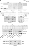
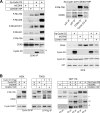
Activating phosphorylations of CDK4 and CDK6 are differentially regulated. (A) Cyclin D3, cyclin D1, or CDK6 coimmunoprecipitates (IP) from serum-stimulated T98G or CEM cells were separated by 2D gel electrophoresis and electroblotted for immunodetection using the phospho-CDK4 (T172) antibody (P-T172), which also recognizes T177-phosphorylated CDK6. The same membranes were then reprobed for detection of total CDK4 and CDK6 by the use of anti-CDK4 and anti-CDK6 antibodies. Double arrows indicate the positions of the forms of CDK4 phosphorylated at T172 and CDK6 phosphorylated at T177. IEF, isoelectric focusing. (B) Extracts from HEK, T98G, or CHO cells transfected using plasmids encoding untagged cyclin D3 or cyclin D3-X-press (Xp-Cyclin D3) with or without CDK4-HA or untagged CDK6 were coimmunoprecipitated using cyclin D3 antibody (upper panel) or using antibodies against HA-tag (CDK4-HA IP), CDK6, cyclin D3, p21 (C-19 from Santa Cruz), or p27 (C-15 from Santa Cruz) or mouse and rabbit control immunoglobulins (IgG) (lower panel). The immunoprecipitates were assayed for pRb kinase activity, separated by SDS-PAGE, and immunoblotted. The pRb-kinase activity shown by in vitro phosphorylation of the pRb fragment at S780 was detected using a phosphospecific antibody (PRb-780). Cyclin D3 and CDK4 or CDK6 (in the appropriate transfections) were detected using specific antibodies. The upper band in the panel depicting the detection of Xp-Cyclin D3 (*) resulted from its phosphorylations by CDK4 during incubation with ATP for the pRb kinase activity assay. (C) The same extracts from transfected HEK, T98G, and CHO cells were coimmunoprecipitated using cyclin D3 antibody, separated by 2D gel electrophoresis, and electroblotted for immunodetection using anti-CDK4 or anti-CDK6 antibody. The positions of T172-phosphorylated CDK4 and T177-phosphorylated CDK6 are indicated by arrows.
FIG. 3.
FIG. 3.
Ectopic cyclin D3, CDK4, and CDK6 and each respective mutant display an overall cellular location that partly differs from the mostly nuclear compartmentalization of endogenous CAK (cyclin H and CDK7). (A) Indirect immunofluorescent codetection of cyclin D3 (green) and either CDK4 or CDK6 (red) from CHO cells transfected using plasmids encoding cyclin D3-X-press and wtCDK4-HA, CDK4P173S-HA, wtCDK6, or CDK6S178P. (B) Indirect immunofluorescent codetection of CDK7 (green) and cyclin H (red) from nontransfected CHO cells. For panels A and B, nuclei were counterstained using Hoechst 33258 dye.
FIG. 4.
FIG. 4.
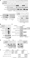
Proline 173 is essential for T172 phosphorylation and activity of CDK4 in intact cells. (A) Extracts from CHO cells transfected using plasmids encoding cyclin D3-X-press (Xp-Cyclin D3) and wtCDK4-HA, CDK4P173S-HA, or CDK4P173H-HA, alone or in combination, were coimmunoprecipitated (IP) using anti-HA (HA IP) or anti-cyclin D3 (D3 IP) antibody or (as controls; lower left panel) using antibodies against p21 or p27 antibodies, assayed for pRb kinase activity, separated by SDS-PAGE, and immunoblotted. The in vitro S780 phosphorylation of the pRb fragment (P-Rb-780) was detected using a phosphospecific antibody. CDK4 and cyclin D3 were also detected. In the lower right panel, whole-cell-extract (WCE) detections of ectopic and endogenous proteins from the same samples are shown. CHO cells endogenously express CDK4 (lower band in CDK4 detection), cyclin D1, and p27 but not cyclin D3. (B) In the same experiment, the in vitro phosphorylation of the pRb fragment was analyzed using phosphospecific antibodies directed against phosphorylated T826 (P-Rb-826), S807 and S811 (P-Rb-807/811), and S807 (P-Rb-807) and S795 (P-Rb-795). (C) The same extracts as those described for panel A were coimmunoprecipitated using HA or cyclin D3 antibody, separated by 2D gel electrophoresis, and electroblotted for immunodetection using anti-CDK4 or the phospho-CDK4 (T172) antibody (P-T172) followed by detection of total CDK4 on the same membrane. (D and E) Extracts from HEK, T98G, or HCT116 cells transfected as described for panel A, or with a plasmid encoding cyclin D1-Flag instead of cyclin D3, were coimmunoprecipitated using anti-cyclin D3 (Cyclin D3 IP or D3 IP) or anti-Flag (D1-Flag IP) antibodies and assayed as described above for pRb kinase activity (P-Rb-780) (D) or separated by 2D gel electrophoresis and electroblotted for immunodetection using anti-CDK4 antibody (E). The upper band in the panels depicting detection of Xp-Cyclin D3 (* in panels A, B, and D) resulted from its in vitro phosphorylations by CDK4 during the pRb kinase activity assay. Arrows in panels C and E indicate the position of the T172-phosphorylated form of CDK4.
FIG. 5.
FIG. 5.
CDK4 proline 173 is essential for mitogenesis. Quiescent T98G cells were transfected with plasmids encoding wt CDK4-HA, T172ACDK4-HA, or P173SCDK4-HA and stimulated with 15% FBS for 16 h with BrdU during the last 30 min. In duplicate dishes, DNA synthesis was evaluated in 100 cells/dish displaying the HA epitope by counting the proportion of nuclei showing incorporation of BrdU. The upper panel illustrates the double immunofluorescence detection of the HA epitope (red) and BrdU (green).
FIG. 6.
FIG. 6.
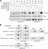
Proline 173 is not essential for in vitro activation of CDK4 by CAK (CDK7). Extracts from CHO cells transfected using plasmids encoding cyclin D3-X-press (Xp-Cyclin D3) and wt CDK4-HA, CDK4P173S-HA, CDK4P173H-HA, or CDK4T172A-HA, alone or in combination, were coimmunoprecipitated (IP) using anti-HA (HA IP) or anti-cyclin D3 (D3 IP) antibody. The immunoprecipitated complexes were incubated without (−) or with (+) recombinant cyclin H-CDK7-Mat1 complex (CAK) and ATP, assayed for their pRb kinase activity (P-Rb-780) as described for Fig. 4A (A), or separated by 2D gel electrophoresis and electroblotted for immunodetection using anti-CDK4 (B). Arrows indicate the position of the T172-phosphorylated form of CDK4.
FIG. 7.
FIG. 7.
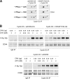
In vitro activation of cyclin D3-bound CDK4, CDK4P173S, and CDK6 by CAK in the presence of different ATP concentrations. Extracts from CHO cells transfected using plasmids encoding cyclin D3-X-press and wt CDK4-HA, CDK4P173S-HA, or wt CDK6 were coimmunoprecipitated (IP) using anti-cyclin D3 antibody. (A) The immunoprecipitated complexes were preincubated without (−) or with (+) λ-phosphatase (PPase), extensively washed, and then incubated without or with 1 μg of recombinant cyclin H-CDK7-Mat1 complex (CAK) and 2 mM ATP, separated by 2D gel electrophoresis, and electroblotted for immunodetection using anti-CDK4. Arrows indicate the position of the T172-phosphorylated form of CDK4. (B) Cyclin D3 complexes (Cyclin D3 IP) or mock immunoprecipitates (IgG IP) processed using the same CHO cell extracts were dephosphorylated by preincubation with λ-phosphatase, extensively washed, and then incubated with 0.25 or 1 μg of CAK in the presence of different ATP concentrations. After thorough washings, the activation of cyclin D3-CDK4/CDK6 complexes was assessed by their pRb-kinase activity (P-Rb-780) codetected with CDK4 or CDK6 (arrowhead).
FIG. 8.
FIG. 8.
S178P mutation activates CDK6. (A) Extracts from CHO cells transfected using plasmids encoding cyclin D3-X-press (Xp-Cyclin D3) and wt CDK6 or CDK6S178P, alone or in combination, were coimmunoprecipitated (IP) using anti-CDK6 antibody or anti-cyclin D3 antibody or (as controls; upper right panel) using antibodies against p21 or p27 antibodies, assayed for pRb kinase activity, separated by SDS-PAGE, and immunoblotted. The in vitro phosphorylation of the pRb fragment was analyzed using phosphospecific antibodies directed against phosphorylated S780 (P-Rb-780), T826 (P-Rb-826), S807 and S811 (P-Rb-807/811), T821 (P-Rb-821), and S795 (P-Rb-795). CDK6 and cyclin D3 were also detected from the same membranes. In the lower right panel, whole-cell-extract (WCE) detections of ectopic and endogenous proteins from experiments using the same samples are shown. (B) Extracts from HEK, T98G, and HCT116 cells, transfected using plasmids encoding cyclin D3-X-press (Xp-Cyclin D3) or cyclin D1-Flag and wtCDK6 or CDK6S178P, alone or in combination, were coimmunoprecipitated using anti-cyclin D3 (Cyclin D3 IP or D3 IP), anti-Flag (D1-Flag IP), or anti-CDK6 (K6 IP) antibody, assayed for pRb kinase activity, separated by SDS-PAGE, and immunoblotted. The in vitro phosphorylation of the pRb fragment (P-Rb-780), CDK6, and cyclin D3 or cyclin D1 (in the appropriate transfections) was then detected using membranes from the same experiment. The upper band in the panels depicting detection of Xp-Cyclin D3 (*) resulted from its in vitro phosphorylation by CDK6S178P during the pRb kinase activity assay.
FIG. 9.
FIG. 9.
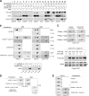
S178P mutation enforces T177 phosphorylation and activity of CDK6. (A to C) Extracts from CHO cells transfected using plasmids encoding cyclin D3-X-press (Xp-Cyclin D3) and wt CDK6 or CDK6S178P, alone or in combination, were coimmunoprecipitated (IP) using anti-CDK6 (CDK6 IP) or anti-cyclin D3 (D3 IP) antibody. The immunoprecipitated complexes were incubated without (−) or with (+) recombinant cyclin H-CDK7-Mat1 complex (CAK) and ATP. (A) The complexes were then assayed for pRb kinase activity, which was detected using the S780 pRb phospho-specific antibody (P-Rb-780). CDK6 and cyclin D3 were then detected on the same membrane. The upper band in the panel depicting detection of Xp-Cyclin D3 (*) resulted from its in vitro phosphorylations by CDK6 during the pRb kinase activity assay. (B, upper panel) The immunoprecipitated complexes incubated without or with CAK as described for panel A were separated by 2D gel electrophoresis and electroblotted for immunodetection using the phospho-CDK4 (T172) antibody (P-T172) (which detects T177-phosphorylated CDK6) or the CDK6 antibody for the detection of total CDK6. (B, lower panel) The same extracts from cells transfected using plasmids encoding wt CDK6 or CDK6S178P were coimmunoprecipitated using anti-cyclin D1 (D1 IP), anti-cyclin D3 (D3 IP), or anti-CDK6 (K6 IP) antibodies, separated by SDS-PAGE, and electroblotted for immunodetection using the CDK6 antibody. (C) The immunoprecipitated complexes were preincubated without (−) or with (+) λ-phosphatase (PPase), extensively washed, and either incubated without or with 1 μg CAK and 2 mM ATP, separated by 2D gel electrophoresis, and electroblotted for immunodetection using anti-CDK6 (upper panel) or incubated with 0.25 or 1 μg CAK in the presence of different ATP concentrations, and the activation of cyclin D3-CDK6S178P was assessed by its pRb-kinase activity (P-Rb-780) codetected with CDK6 (lower panel). (D) Extracts from HEK and T98G cells transfected using plasmids encoding cyclin D3-X-press or cyclin D1-Flag and wt CDK6 or CDK6S178P were coimmunoprecipitated using cyclin D3 or Flag (D1-Flag IP) antibodies, separated by 2D gel electrophoresis, and electroblotted for immunodetection using anti-CDK6 antibody. (E) Extracts from CHO cells transfected using plasmids encoding wt CDK6 or CDK6S178H and cyclin D3-X-press (Cyclin D3) were coimmunoprecipitated using anti-CDK6 or anti-cyclin D3 antibody, separated by 2D gel electrophoresis, and electroblotted for immunodetection using the CDK6 antibody. Arrows indicate the position of the T177-phosphorylated form of CDK6.

References

    1. Aprelikova, O., Y. Xiong, and E. T. Liu. 1995. Both p16 and p21 families of cyclin-dependent kinase (CDK) inhibitors block the phosphorylation of cyclin-dependent kinases by the CDK-activating kinase. J. Biol. Chem. 27018195-18197. - PubMed
    1. Bagui, T. K., S. Mohapatra, E. Haura, and W. J. Pledger. 2003. P27Kip1 and p21Cip1 are not required for the formation of active D cyclin-cdk4 complexes. Mol. Cell. Biol. 237285-7290. - PMC - PubMed
    1. Bartek, J., J. Bartkova, and J. Lukas. 1996. The retinoblastoma protein pathway and the restriction point. Curr. Opin. Cell Biol. 8805-814. - PubMed
    1. Bartkova, J., J. Lukas, M. Strauss, and J. Bartek. 1998. Cyclin D3: requirement for G1/S transition and high abundance in quiescent tissues suggest a dual role in proliferation and differentiation. Oncogene 171027-1037. - PubMed
    1. Berthet, C., K. D. Klarmann, M. B. Hilton, H. C. Suh, J. R. Keller, H. Kiyokawa, and P. Kaldis. 2006. Combined loss of Cdk2 and Cdk4 results in embryonic lethality and Rb hypophosphorylation. Dev. Cell 10563-573. - PubMed

Publication types

MeSH terms

LinkOut - more resources