. 2009 Aug;29(15):4201-19.
doi: 10.1128/MCB.00056-09. Epub 2009 Jun 1.

ADAMTS-7, a direct target of PTHrP, adversely regulates endochondral bone growth by associating with and inactivating GEP growth factor

Affiliations

ADAMTS-7, a direct target of PTHrP, adversely regulates endochondral bone growth by associating with and inactivating GEP growth factor

Xiao-Hui Bai et al. Mol Cell Biol. 2009 Aug.

Abstract

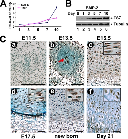
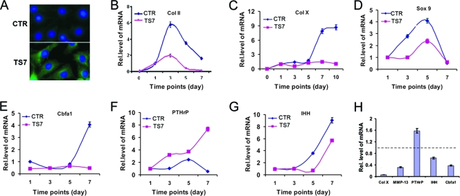
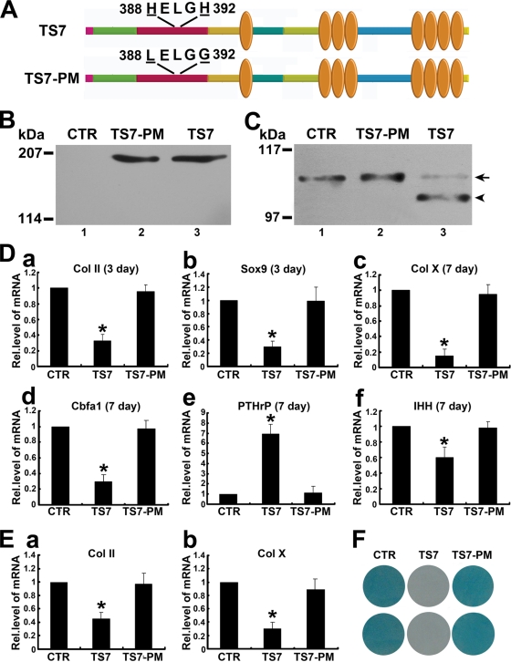
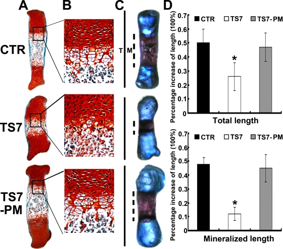
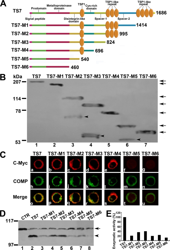
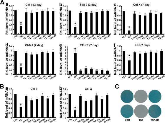
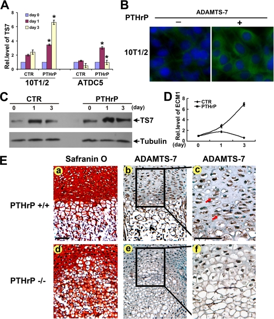
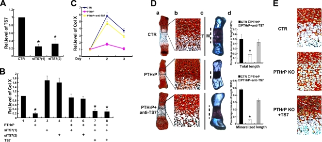
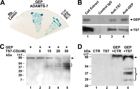
ADAMTS-7, a metalloproteinase that belongs to ADAMTS family, is important for the degradation of cartilage extracellular matrix proteins in arthritis. Herein we report that ADAMTS-7 is upregulated during chondrocyte differentiation and demonstrates the temporal and spatial expression pattern during skeletal development. ADAMTS-7 potently inhibits chondrocyte differentiation and endochondral bone formation, and this inhibition depends on its proteolytic activity. The cysteine-rich domain of ADAMTS-7 is required for its interaction with the extracellular matrix, and the C-terminal four-thrombospondin motifs are necessary for its full proteolytic activity and inhibition of chondrocyte differentiation. ADAMTS-7 is an important target of canonical PTHrP signaling, since (i) PTHrP induces ADAMTS-7, (ii) ADAMTS-7 is downregulated in PTHrP null mutant (PTHrP-/-) growth plate chondrocytes, and (iii) blockage of ADAMTS-7 almost abolishes PTHrP-mediated inhibition of chondrocyte hypertrophy and endochondral bone growth. ADAMTS-7 associates with granulin-epithelin precursor (GEP), an autocrine growth factor that has been implicated in tissue regeneration, tumorigenesis, and inflammation. In addition, ADAMTS-7 acts as a new GEP convertase and neutralizes GEP-stimulated endochondral bone formation. Collectively, these findings demonstrate that ADAMTS-7, a direct target of PTHrP signaling, negatively regulates endochondral bone formation by associating with and inactivating GEP chondrogenic growth factor.


Figures

FIG. 1.
FIG. 1.
Expression of ADAMTS-7 in the course of chondrogenesis in vitro and in the growth plate chondrocytes in vivo. (A) Expression of ADAMTS-7 and collagen X was examined in the course of chondrogenesis of a micromass culture of C3H10T1/2 cells. Micromass cultures of C3H10T1/2 cells were stimulated with BMP-2 protein for various time points as indicated, and the mRNA levels of ADAMTS-7 (TS7) and Col X were assayed using real-time PCR. Rel.level, relative level. (B) Differential expression of ADAMTS-7 protein during chondrogenesis of a micromass culture of C3H10T1/2 cells. C3H10T1/2 cells were cultured as described for panel A, and the levels of ADAMTS-7 (TS-7) and tubulin (serving as an internal control) were detected by using immunoblotting. (C) Temporal and spatial expression of ADAMTS-7 during chondrogenesis in vivo, assayed by immunohistochemistry. The sections of long bone from various embryonic and postnatal developmental stages of mice, as indicated, were stained with anti-ADAMTS7 antiserum (brown) and counterstained with methyl green (green). Immunostaining revealed positive cytoplasmic staining in chondrocytes (insets). Bar = 100 μm.
FIG. 2.
FIG. 2.
ADAMTS-7 is a negative regulator of chondrocyte differentiation. (A) Overexpression of ADAMTS-7 in the ADAMTS-7 stable line. Both control (CTR) (top) and ADAMTS-7 (TS7) (bottom) cell lines based on C3H10T1/2 cells were stained with anti-ADAMTS-7 antibodies. The nuclei were stained with 4′,6-diamidino-2-phenylindole (DAPI). (B to G) Overexpression of ADAMTS-7 regulates the expression of genes critical for chondrogenesis. Micromass cultures of both control and ADAMTS-7 stable lines were stimulated with BMP-2 protein for various time points as indicated, and the levels of mRNAs were determined using real-time PCR. (H) Effect of ADAMTS-7-conditioned medium on the expression of molecules controlling chondrocyte maturation. Micromass cultures of C3H10T1/2 cells were cultured in the presence of either control or ADAMTS-7-conditioned medium for 7 days. The units are arbitrary, and the normalized values were calibrated against controls, here given the value of 1. Rel.level, relative level.
FIG. 3.
FIG. 3.
The proteolytic activity of ADAMTS-7 is required for its regulation of chondrogenesis. (A) Schematic structure of the ADAMTS-7 and its point mutant. HELGH in the catalytic domain was substituted by LELGG, as indicated. (B) Western blot analysis of ADAMTS-7 and its point mutant. The HEK 293-EBNA cells were transfected with either control (CTR) (lane 1) or expression constructs encoding wild-type ADAMTS-7 (TS7) (lane 3) or ADAMTS-7 with mutant catalytic domain (TS7-PM) (lane 2). The conditioned medium was subjected to reduced 8% SDS-PAGE and detected with polyclonal anti-ADAMTS-7 antibodies. (C) In vitro digestion assay of COMP by ADAMTS-7 and its point mutant. COMP was incubated with the conditioned medium collected from HEK 293-EBNA cells stably transfected by pcDNA3.1 vector (CTR) (lane 1) or expression constructs encoding wild-type ADAMTS-7 (TS7) (lane 3) or an ADAMTS-7 point mutant (TS7-PM) (lane 2), as indicated. The cleaved products were subjected to reduced 8% SDS-PAGE and detected with polyclonal anti-COMP antibodies. The intact COMP and its digested fragments are indicated with an arrow and arrowhead, respectively. (D) ADAMTS-7 inhibits chondrocyte differentiation, whereas the point mutant of ADAMTS-7 lacking enzymatic activity loses this inhibition. Micromass cultures of C3H10T1/2 cells stably transfected with a control (CTR), ADAMTS-7 (TS7), and its point mutant (TS7-PM) expression plasmid were incubated with 300 ng/ml of BMP-2, and the mRNA levels of Col II (a), Sox9 (b), Col X (c), Cbfa1 (d), PTHrP (e), and IHH (f) were determined by using real-time PCR. The units are arbitrary, and the normalized values were calibrated against the control (CTR), here given the value of 1. Asterisks indicate a significant increase or decrease compared to the control (P < 0.05). (E) Effect of ADAMTS-7 and its point mutant on chondrocyte differentiation using human MSCs. Human MSCs pellets were incubated with BMP-2 for 14 days in the presence of the conditioned medium obtained from either control (CTR), ADAMTS-7 (TS7), or its point mutant (TS7-PM) stably transfected HEK 293-EBNA cell lines, and Col II (a) and Col X (b) expression was analyzed by real-time PCR. (F) Whole-mount alcian blue histochemistry. Staining was performed on the micromass culture of pcDNA3.1 (CTR), ADAMTS-7 point mutant (TS7-PM), and wild-type ADAMTS-7 (TS7) stable lines based on C3H10T1/2 cells treated with BMP-2 protein for 14 days. Rel.level, relative level.
FIG. 4.
FIG. 4.
ADAMTS-7-mediated chondrocyte hypertrophy, mineralization, and bone length depends on its enzymatic activity. (A and B) Safranin O-fast green staining of metatarsals. Metatarsals were explanted from 15-day-old mouse embryos and cultured in the presence of conditioned medium of control (CTR), ADAMTS-7 (TS7), or its point mutant (TS7-PM). After 5 days of culture, the explants were fixed and safranin O-fast green staining was observed in low-power (A) or high-power (B) microphotography. (C) Alizarin red S and alcian blue staining of metatarsals. The explants were fixed and processed for alizarin red S and alcian blue staining, and a representative photograph of an explanted metatarsal after 5 days of culture is presented. (D) Percent increase in total and mineralization length of metatarsal bones. Metatarsals were cultured as described above, total length or mineralization length was determined, and the percent increase was calculated (percent increase = [length at day 5 − length at day 0]/length at day 0). Asterisks indicate a significant difference from the control (P < 0.05).
FIG. 5.
FIG. 5.
Effects of ADAMTS-7 functional domains on its cell surface location and enzymatic activity. (A) Schematic structure of the ADAMTS-7 and its C-terminal deletion mutants. Numbers refer to amino acid residues in ADAMTS-7. The functional domains are indicated. (B) Western blotting analysis of ADAMTS-7 and its C-terminal deletion mutants. The conditioned medium collected from HEK 293-ENBA cells stably transfected by either ADAMTS-7 or its C-terminal deletion mutants was detected by Western blotting analysis with anti-c-Myc antibodies. The arrows indicate the full length of the ADAMTS-7 and its series of mutants, and the arrowheads indicate the fragments resulted from several ADAMTS-7 deletion mutants. (C) The subcellular localization of the ADAMTS-7 and its domain deletion mutants in RCS cells. RCS chondrocytes were transiently transfected with expression constructs encoding the ADAMTS-7 and its C-terminal deletion mutants. The expression of ADAMTS-7 or its C-terminal deletion mutants was visualized with anti-c-Myc antibodies. COMP was visualized by cell staining with polyclonal anti-COMP antibodies. Overlapping signals are indicated with “Merge.” (D) In vitro digestion assays of COMP mediated by ADAMTS-7 and its C-terminal domain deletion mutants. Purified COMP (200 nM) was incubated with the conditioned medium collected from HEK 293-EBNA cells stably transfected by pcDNA3.1 (CTR), ADAMTS-7 (TS7), or its C-terminal domain deletion mutants (TS7-M1 to TS7-M6), as indicated. The cleaved products were subjected to reduced 8% SDS-PAGE and detected with polyclonal anti-COMP antibodies. The intact COMP and its digested fragments are indicated with an arrow and arrowhead, respectively. (E) Qualitative analysis of enzymatic activity of ADAMTS-7 and its C-terminal domain deletion mutants. The values were calibrated against ADAMTS-7 (TS7), here given the value of 100%.
FIG. 6.
FIG. 6.
Substrate-binding domain of ADAMTS-7 is important for its regulation of chondrogenesis. (A) Effect of ADAMTS-7 and its C-terminal deletion mutants on chondrocyte differentiation of murine C3H10T1/2 cells. Micromass cultures of control (CTR), wild-type ADAMTS-7 (TS7), or its C-terminal mutants (TS7-M1 to TS7-M6) C3H10T1/2 stable cell lines were stimulated with 300 ng/ml BMP-2 protein for 3 or 7 days, and the mRNA levels of Col II (a), Sox9 (b), Col X (c), Cbfa1 (d), PTHrP (e), and IHH (f) were determined by using real-time PCR. The units are arbitrary, and the normalized values were calibrated against the control (CTR), here given the value of 1. Asterisks indicate a significant increase or decrease from the control (P < 0.05). (B) Effect of ADAMTS-7 and its C-terminal deletion mutants on the expressions of Col II and Col X during chondrogenesis of human MSCs. Human MSCs pellets were cultured for 14 days in the presence of the conditioned medium obtained from the control (CTR), ADAMTS-7 (TS7), or its C-terminal mutants (TS7-M1 to TS7-M6) stably transfected HEK 293-EBNA cell lines, and Col II (a) and Col X (b) expression was analyzed by real-time PCR as described for panel A. (C) Whole-mount alcian blue histochemistry. Staining was performed on the micromass culture of pcDNA3.1 (CTR), wild-type ADAMTS-7 (TS7), and C-terminal four-thrombospondin motifs deletion mutant (TS7-M1) stable lines based on C3H10T1/2 cells treated with BMP-2 protein for 14 days. Rel.level, relative level.
FIG. 7.
FIG. 7.
ADAMTS-7 expression depends on PTHrP signaling. (A) PTHrP induces the expression of ADAMTS-7 mRNA, assayed by real-time PCR. 10T1/2 and ATDC5 cells pretreated with recombinant 300 ng/ml of BMP-2 for 1 week were cultured without PTHrP (CTR) or with PTHrP (10−7 M) for various time periods, as indicated. The normalized values against GAPDH were calibrated against controls (day 0), given the value of 1. Asterisk indicates a significant difference from the control at corresponding time points (P < 0.05). (B) PTHrP increases the level of ADAMTS-7 protein, assayed by immunofluorescent cell staining. Micromass cultures of C3H10T1/2 cells treated with or without PTHrP (10−7 M) for 3 days were stained with anti-ADAMTS-7 antibodies (green). The nuclei were stained with 4′,6-diamidino-2-phenylindole (DAPI). (C) PTHrP induces the expression of ADAMTS-7 protein, assayed by Western blotting. ATDC5 cells pretreated with recombinant 300 ng/ml of BMP-2 for 1 week were cultured without PTHrP (CTR) or with PTHrP (10−7 M) for various time periods, as indicated. The cell lysates were detected with either anti-ADAMTS-7 or antitubulin (serving as an internal control) antibodies. (D) PTHrP induces the expression of ECM1 mRNA, assayed by real-time PCR. 10T1/2 cells pretreated with recombinant 300 ng/ml of BMP-2 for 1 week were cultured without PTHrP (CTR) or with PTHrP (10−7 M) for various time periods, as indicated. The normalized values against GAPDH were calibrated against controls (day 0), given the value of 1. (E) ADAMTS-7 is markedly reduced in the growth plate chondrocytes of PTHrP null mice, revealed by immunohistochemistry. Safranin O staining was performed using sections of long bone from 18.5-day-old wild-type (a) and PTHrP null mutant (d) mouse embryos. (b and e) Low-power microphotograph of section stained with anti-ADAMTS-7 antibodies (brown, indicated with arrows) and counterstained with methyl green (green). (c and f) High-power microphotograph of the section shown in panels b and e. Rel.level, relative level. Bar = 100 μm.
FIG. 8.
FIG. 8.
ADAMTS-7 is required for the PTHrP-mediated inhibition on chondrocyte differentiation and endochondral bone formation. (A) ADAMTS-7 siRNAs efficiently repress the expression of endogenous ADAMTS-7 in C3H10T1/2 cells. C3H10T1/2 cells were transfected with either pSuper vector (CTR) or one of two ADAMTS-7 siRNAs cloned into pSuper, as indicated, and the level of ADAMTS-7 was measured using real-time PCR. (B) Knockdown of ADAMTS-7 almost abolished PTHrP-mediated inhibition of Col X expression, whereas its reexpression largely restored PTHrP action. Micromass cultures of C3H10T1/2 cells transfected with pSuper, siTS7 [siTS7(1), siTS7(2)], ADAMTS-7 (TS7) expression plasmid, or various combinations as indicated were pretreated with BMP-2 and then cultured in the presence or absence of PTHrP (10−7 M), and the level of Col X was determined by real-time PCR. The units are arbitrary, and the normalized values were calibrated against the control, here given the value of 1. (C) ADAMTS-7 antibody dramatically attenuated PTHrP-mediated inhibition of Col X. ATDC5 cells pretreated with BMP-2 for 3 days were cultured in the absence (CTR) or presence of PTHrP (10−7M) without (PTHrP) or with anti-ADAMTS-7 (1 μg/ml) antibodies (PTHrP + anti-TS7) for various time points as indicated, and Col X was measured and analyzed as described for panel B. (D) ADAMTS-7 blocking antibody completely neutralized PTHrP-mediated chondrocyte hypertrophy, mineralization, and bone length. (a and b) Safranin O-fast green staining of metatarsals. Metatarsals were explanted from 15-day-old mouse embryos and cultured in the absence (CTR) or presence of PTHrP (10−7 M) with or without anti-ADAMTS7 (1 μg/ml) antibodies. The explants were cultured for 5 days, and safranin O-fast green staining was observed by using low-power (a) or high-power (b) microphotography. (c) Alizarin red S and alcian blue staining of metatarsals. Metatarsals were cultured as described above and processed for alizarin red S and alcian blue staining; a representative photograph is presented. (d) Percent increase in total and mineralization length of metatarsal bones. Metatarsals were cultured as described above, total or mineralization length was determined, and the percent increase was calculated (percent increase = [length at day 5 − length at day 0]/length at day 0). Asterisk indicates a significant difference from the control (P < 0.05). (E) ADAMTS-7 corrects the defects in chondrocyte hypertrophy in the metatarsal bones of PTHrP null embryos. Metatarsals were explanted from 14.5-day-old wild-type and PTHrP−/− mouse embryos and cultured in the absence (CTR) or presence of ADAMTS-7-conditioned medium. The explants were cultured for 5 days, and safranin O-fast green staining was observed. Rel.level, relative level.
FIG. 9.
FIG. 9.
ADAMTS-7 associates with and digests GEP. (A) ADAMTS-7 binds to GEP in yeast. Yeast two-hybrid assay to test the interaction of proteins fused to the VP16 AD and proteins fused to the Gal4 DNA binding domain. Each pair of plasmids, as indicated, encoding proteins fused to VP16 (below the line) in the vector pPC86 (i.e., pPC86-c-jun, pPC86-GEP, and pPC86-Rb) and those encoding proteins fused to Gal4 (above the line) in the vector pDBleu (i.e., pDB-c-fos, pDB-ADAMTS-7, and pDB-lamin) were cotransfected into yeast strain MAV203. Yeast transformants were selected on SD-Leu-Trp plates and tested for β-galactosidase activity. The known interaction between c-Jun and c-Fos was used as a positive control, whereas the lack of interaction between Rb and lamin was used as a negative control. (B) ADAMTS-7 binds to GEP in chondrocytes (Co-IP assay). Cell extracts prepared from human chondrocytes were incubated with anti-GEP antibodies, anti-ADAMTS-7 antibodies, or control IgG. The immunoprecipitated protein complex and cell extracts (lane 1, which provides a positive control) were examined by Western blotting with either anti-GEP (top) or anti-ADAMTS-7 (bottom) antibodies. (C) The catalytic domain of ADAMTS-7 digests GEP in a dose-dependent manner. GEP was incubated with various amounts of catalytic domain of ADAMTS-7 (TS7-CD), as indicated; the cleaved products were separated by nonreduced SDS-PAGE, and intact GEP and fragments were detected with polyclonal anti-GEP antibodies. The intact GEP (arrow) and processed fragments are indicated. (D) Intact ADAMTS-7 cleaves GEP. GEP was incubated with either the control (CTR) (lane 3) or ADAMTS-7-conditioned medium (lane 4) for 4 h. The processed products as well as the control and ADAMTS-7 media were detected with anti-GEP antibodies. The intact GEP (arrow) and processed fragments are indicated.
FIG. 10.
FIG. 10.
ADAMTS-7 inactivates the chondroinductive action of GEP. (A) ADAMTS-7 inhibits GEP-induced Col X and MMP-13 expression, assayed by real-time PCR. ATDC5 cells pretreated with BMP-2 for 5 days were cultured in the absence or presence of GEP, ADAMTS-7 (TS7)-, ADAMTS-7 point mutant (TS7PM)-, or granulin B (GRN B)-conditioned medium or various combinations as indicated for an additional 3 days, and Col X and MMP-13 expression was measured by real-time PCR. The units are arbitrary, and the normalized values were calibrated against controls, here given the value of 1. Asterisks indicate a significant difference from the control (P < 0.05). (B) ADAMTS-7 abolishes GEP-stimulated endochondral ossification. (a and b) Safranin O-fast green staining of metatarsals. Metatarsals were explanted from 15-day-old mouse embryos and cultured in the absence (CTR) or presence of GEP with or without ADAMTS7-conditioned medium. After 5 days of culture, safranin O-fast green staining was observed by using low-power (a) or high-power (b) microphotography. (c) Alizarin red S and alcian blue staining of metatarsals. The explants were fixed and processed for alizarin red S and alcian blue staining; a representative photograph is presented. (d) Percent increase in total and mineralization length of metatarsal bones. Metatarsals were cultured as described above, the total or mineralization length was determined, and the percent increase was calculated (percent increase = [length at day 5 − length at day 0]/length at day 0). Asterisks indicate a significant difference from the control (P < 0.05). (C) Proposed model for explaining the role and regulation of ADAMTS-7 in chondrocyte differentiation. ADAMTS-7 metalloproteinase, a direct target of PTHrP, negatively regulates chondrocyte differentiation by associating with GEP and inactivating chondrogenic activity of GEP growth factor. Rel.level, relative level.

References

    1. Abbaszade, I., R. Q. Liu, F. Yang, S. A. Rosenfeld, O. H. Ross, J. R. Link, D. M. Ellis, M. D. Tortorella, M. A. Pratta, J. M. Hollis, R. Wynn, J. L. Duke, H. J. George, M. C. Hillman, Jr., K. Murphy, B. H. Wiswall, R. A. Copeland, C. P. Decicco, R. Bruckner, H. Nagase, Y. Itoh, R. C. Newton, R. L. Magolda, J. M. Trzaskos, T. C. Burn, et al. 1999. Cloning and characterization of ADAMTS11, an aggrecanase from the ADAMTS family. J. Biol. Chem. 27423443-23450. - PubMed
    1. Amizuka, N., H. Warshawsky, J. E. Henderson, D. Goltzman, and A. C. Karaplis. 1994. Parathyroid hormone-related peptide-depleted mice show abnormal epiphyseal cartilage development and altered endochondral bone formation. J. Cell Biol. 1261611-1623. - PMC - PubMed
    1. Anakwe, O. O., and G. L. Gerton. 1990. Acrosome biogenesis begins during meiosis: evidence from the synthesis and distribution of an acrosomal glycoprotein, acrogranin, during guinea pig spermatogenesis. Biol. Reprod. 42317-328. - PubMed
    1. Arikawa-Hirasawa, E., H. Watanabe, H. Takami, J. R. Hassell, and Y. Yamada. 1999. Perlecan is essential for cartilage and cephalic development. Nat. Genet. 23354-358. - PubMed
    1. Atkinson, B. L., K. S. Fantle, J. J. Benedict, W. E. Huffer, and A. Gutierrez-Hartmann. 1997. Combination of osteoinductive bone proteins differentiates mesenchymal C3H/10T1/2 cells specifically to the cartilage lineage. J. Cell. Biochem. 65325-339. - PubMed

Publication types

MeSH terms