. 2009 Jun 1;23(11):1303-12.
doi: 10.1101/gad.1803909.

L1 retrotransposition occurs mainly in embryogenesis and creates somatic mosaicism

Affiliations

L1 retrotransposition occurs mainly in embryogenesis and creates somatic mosaicism

Hiroki Kano et al. Genes Dev. .

Abstract

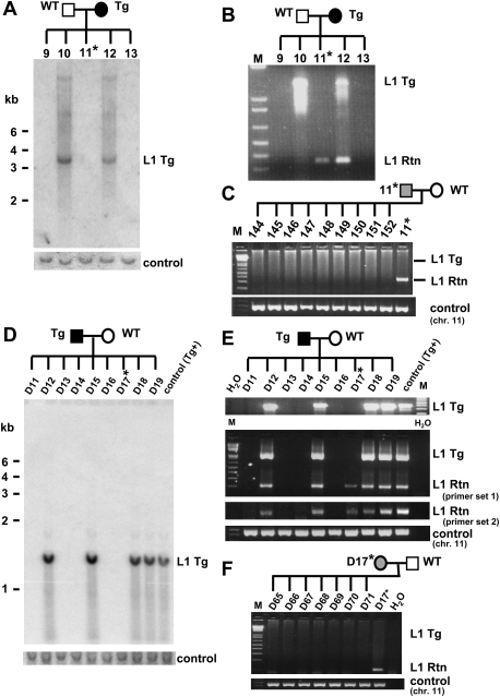
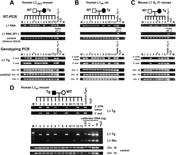
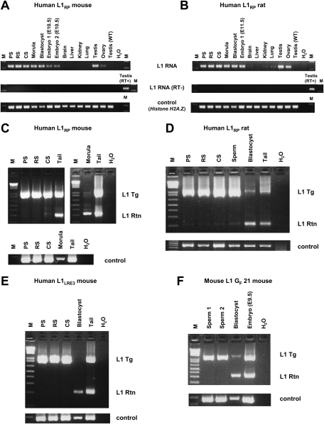
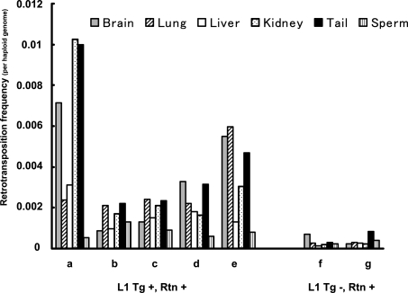
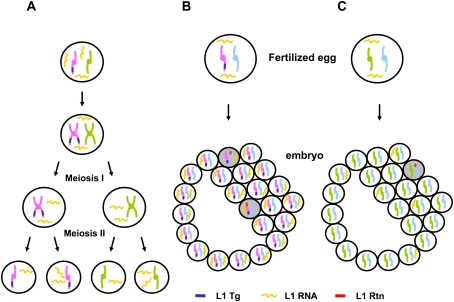
Long Interspersed Element 1 (L1) is a retrotransposon that comprises approximately 17% of the human genome. Despite its abundance in mammalian genomes, relatively little is understood about L1 retrotransposition in vivo. To study the timing and tissue specificity of retrotransposition, we created transgenic mouse and rat models containing human or mouse L1 elements controlled by their endogenous promoters. Here, we demonstrate abundant L1 RNA in both germ cells and embryos. However, the integration events usually occur in embryogenesis rather than in germ cells and are not heritable. We further demonstrate L1 RNA in preimplantation embryos lacking the L1 transgene and L1 somatic retrotransposition events in blastocysts and adults lacking the transgene. Together, these data indicate that L1 RNA transcribed in male or female germ cells can be carried over through fertilization and integrate during embryogenesis, an interesting example of heritability of RNA independent of its encoding DNA. Thus, L1 creates somatic mosaicism during mammalian development, suggesting a role for L1 in carcinogenesis and other disease.


Figures

Figure 1.
Figure 1.
L1 retrotransposition caused by L1 RNA carried over through meiosis, fertilization, and embryogenesis in the L1RP mouse (AC) and in the L1LRE3 mouse (DF). (A) Southern blot analysis on tail DNA isolated from offspring of an L1RP transgenic female mouse. A 1.4-kb DNA probe generated from the retrotransposition cassette of the L1RP transgene, which was expected to hybridize to both the transgene and retrotransposition insertion, exhibited only transgene bands in spite of the presence of retrotransposon amplicons by PCR. The membrane was rehybridized with an unrelated DNA probe generated from mouse chromosome 11 as a DNA loading control. (B) Genotyping PCR on tail DNA indicates an L1 retrotransposition event in a mouse lacking the transgene (mouse 11). (C) Mouse 11 (transgene-negative, retrotransposition event-positive) was bred with a wild-type mouse, and its offspring were genotyped. No offspring of this mouse inherited the retrotransposition insertion, indicating mosaicism of the L1 retrotransposition event in mouse 11. A control PCR on mouse chromosome 11 was performed to confirm the amount and quality of DNA. (DF) Similar data to those in A–C are shown for the offspring of an L1LRE3 transgenic male mouse using tail DNA. The transgenic male mouse was bred with a wild-type female mouse, and its offspring were genotyped by Southern blot using a 503-bp probe generated from the L1 3′UTR and SV40 poly(A) signal sequence of the L1LRE3 transgene (D), and by PCR (E). Two independent PCR primer sets were used to confirm the presence of retrotransposition events. (F) The single offspring (D17) that had a retrotransposition event while lacking the L1 transgene was bred with a wild-type mouse. As shown in C, none of its offspring inherited the retrotransposition event. Asterisk denotes a transgene-negative, retrotransposition event-positive mouse. (Tg) transgene; (Rtn) retrotransposition event; (WT) wild-type animal; (M) 1-kb plus DNA Ladder (Invitrogen).
Figure 2.
Figure 2.
Single preimplantation embryos lacking the transgene contain L1 RNA (A–C) and L1 retrotransposition events (D). RT–PCR analysis and genotyping PCR on offspring of an L1LRE3 mouse (A), an L1RP rat (B), and an L1 GF 21 mouse (C). RNA isolated from single morulae or blastocysts was subjected to RT–PCR to detect L1 RNA from the L1 transgene. For genotyping, DNA from each embryo was subjected to genotyping PCR. To exclude the possibility of a false negative genotype for the transgene, each embryo was genotyped by two independent primer sets for the L1 transgene, and three independent genomic loci were amplified as controls. In AC, an asterisk denotes a transgene-negative, L1 RNA-positive preimplantation embryo. (D) Retrotransposition events in individual late blastocysts. Single blastocysts of the L1RP mouse were subjected to genotyping PCR. For semiquantification, mouse DNA that carries one retrotransposition event per diploid genome (Ostertag et al. 2002) was used as a calibrator DNA. (Middle) The amount of DNA of each blastocyst used in the intron-flanking PCR was presumed to be 0.1–0.5 ng. Thus, retrotransposition events appear to be present in much less than one copy per cell. (RT) Reverse transcriptase; (Tg) transgene; (Rtn) retrotransposition event; (WT) wild-type animal; (M) 1-kb plus DNA Ladder (Invitrogen).
Figure 3.
Figure 3.
L1 transcripts and retrotransposition events in various developmental stages and adult tissues. (A,B) RT–PCR on transgenic L1RP mouse (A) and L1RP rat (B) spermatogenic cell fractions, preimplantation embryos (morulae and blastocysts), E10.5–E11.5 embryos, and adult tissues. Only a head portion of E10.5–E11.5 embryos was subjected to RT–PCR in order to eliminate contamination of germ cells in the embryonic developmental stages. Testis from wild-type adult animals was used as a negative control for L1 RNA from the transgene. Histone H2A.Z gene was used as an endogenous control. (C–F) Genotyping PCR on L1RP mouse (C), L1RP rat (D), L1LRE3 mouse (E), and L1 GF 21 mouse (F) spermatogenic cell fractions and pooled preimplantation embryos (L1RP mouse line, 10 morulae; L1RP rat line, 25 blastocysts; L1LRE3 mouse line, nine blastocysts; L1 GF 21 mouse line, 12 blastocysts). Spermatogenic cell fractions were prepared from transgene-positive, retrotransposition event-negative mice (C,E,F) and transgene-positive, retrotransposition event-positive rats (D). Nested PCR was performed on each sample, which was optimized to amplify small products preferentially. In D, similar amounts of DNA (5 ng) from rat spermatogenic cell fractions, pooled blastocysts, and tail were subjected to PCR. Genomic DNA of the Actb region was amplified to confirm the amount of DNA. (M) 1-kb plus DNA Ladder (Invitrogen); (RT) reverse transcriptase; (Tg) transgene; (Rtn) retrotransposition event; (WT) wild type.
Figure 4.
Figure 4.
L1 retrotransposition frequency in the L1RP mouse. DNA derived from brain, lung, liver, kidney, tail, and sperm of transgene-positive, retrotransposition event-positive mice (A–E) and transgene-negative, retrotransposition event-positive mice (F,G) was analyzed by qPCR. Genomic DNA of each tissue was subjected to the intron-flanking PCR reactions and internal control PCR. Both PCR reactions were normalized to those of the calibrator DNA. The calibrator DNA was obtained from a mouse with a germline L1 retrotransposition event identified in our previous studies (Ostertag et al. 2002), which had 0.5 retrotransposition insertions per haploid genome. (Tg) L1 transgene; (Rtn) retrotransposition event.
Figure 5.
Figure 5.
L1 transcripts and L1 retrotransposition in germ cells and early development. (A) Abundant L1 RNA is present in both developing germ cells and meiotic cells. Some L1 RNA segregates from its encoding DNA. However, L1 retrotransposition events are rare in these cells. (B) L1 transgene-positive gametes can be fertilized, carrying L1 RNA into the fertilized egg. L1 RNA transcribed either during germ cell development or during embryogenesis can retrotranspose into the genome. (C) L1 transgene-negative gametes (both sperm and egg) can carryover L1 RNA that has segregated from its encoding DNA into the fertilized egg. This L1 RNA can be the source of infrequent L1 retrotransposition events during embryogenesis. Retrotransposition events in scenario B occur more frequently than those in scenario C (see Fig. 4). (Tg) L1 transgene; (Rtn) retrotransposition event. Shaded cells represent retrotransposition-positive cells.

References

    1. An W, Han JS, Wheelan SJ, Davis ES, Coombes CE, Ye P, Triplett C, Boeke JD. Active retrotransposition by a synthetic L1 element in mice. Proc Natl Acad Sci. 2006;103:18662–18667. - PMC - PubMed
    1. Babushok DV, Ostertag EM, Courtney CE, Choi JM, Kazazian HH., Jr L1 integration in a transgenic mouse model. Genome Res. 2006;16:240–250. - PMC - PubMed
    1. Bourc'his D, Bestor TH. Meiotic catastrophe and retrotransposon reactivation in male germ cells lacking Dnmt3L. Nature. 2004;431:96–99. - PubMed
    1. Branciforte D, Martin SL. Developmental and cell type specificity of LINE-1 expression in mouse testis: Implications for transposition. Mol Cell Biol. 1994;14:2584–2592. - PMC - PubMed
    1. Brouha B, Meischl C, Ostertag E, de Boer M, Zhang Y, Neijens H, Roos D, Kazazian HH., Jr Evidence consistent with human L1 retrotransposition in maternal meiosis I. Am J Hum Genet. 2002;71:327–336. - PMC - PubMed

Publication types

MeSH terms