

Longitudinal pattern of regional brain volume change differentiates normal aging from MCI
- PMID: 19487648
- PMCID: PMC2690968
- DOI: 10.1212/WNL.0b013e3181a82634
Longitudinal pattern of regional brain volume change differentiates normal aging from MCI
Abstract
Background: Neuroimaging measures have potential as surrogate markers of disease through identification of consistent features that occur prior to clinical symptoms. Despite numerous investigations, especially in relation to the transition to clinical impairment, the regional pattern of brain changes in clinically normal older adults has not been established. We predict that the regions that show early pathologic changes in association with Alzheimer disease will show accelerated volume loss in mild cognitive impairment (MCI) compared to normal aging.
Methods: Through the Baltimore Longitudinal Study of Aging, we prospectively evaluated 138 nondemented individuals (age 64-86 years) annually for up to 10 consecutive years. Eighteen participants were diagnosed with MCI over the course of the study. Mixed-effects regression was used to compare regional brain volume trajectories of clinically normal individuals to those with MCI based on a total of 1,017 observations.
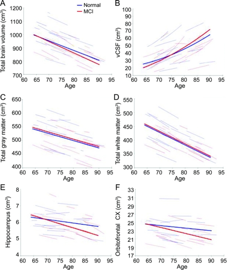
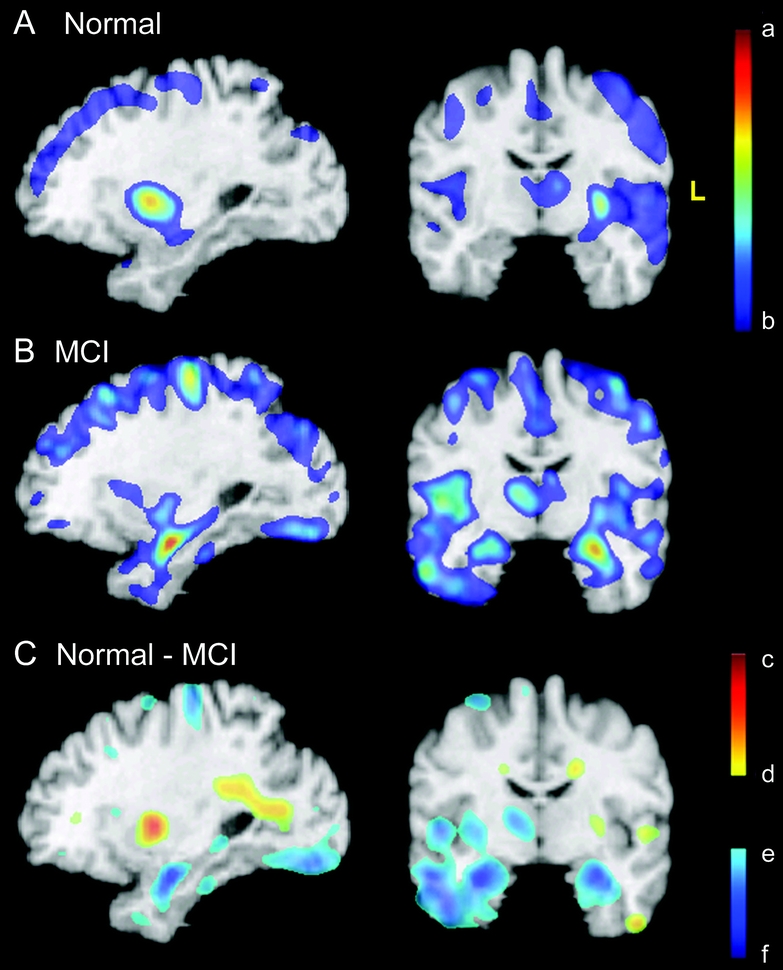
Results: All investigated volumes declined with normal aging (p < 0.05). Accelerated change with age was observed for ventricular CSF (vCSF), frontal gray matter, superior, middle, and medial frontal, and superior parietal regions (p < or = 0.04). The MCI group showed accelerated changes compared to normal controls in whole brain volume, vCSF, temporal gray matter, and orbitofrontal and temporal association cortices, including the hippocampus (p < or = 0.04).
Conclusion: Although age-related regional volume loss is apparent and widespread in nondemented individuals, mild cognitive impairment is associated with a unique pattern of structural vulnerability reflected in differential volume loss in specific regions. Early identification of patterns of abnormality is of fundamental importance for detecting disease onset and tracking progression.
Figures
Similar articles
-
Structural magnetic resonance imaging for the early diagnosis of dementia due to Alzheimer's disease in people with mild cognitive impairment.Cochrane Database Syst Rev. 2020 Mar 2;3(3):CD009628. doi: 10.1002/14651858.CD009628.pub2. Cochrane Database Syst Rev. 2020. PMID: 32119112 Free PMC article.
-
Baseline predictors of rates of hippocampal atrophy in mild cognitive impairment.Neurology. 2007 Oct 9;69(15):1491-7. doi: 10.1212/01.wnl.0000277458.26846.96. Neurology. 2007. PMID: 17923611 Clinical Trial.
-
Longitudinal progression of Alzheimer's-like patterns of atrophy in normal older adults: the SPARE-AD index.Brain. 2009 Aug;132(Pt 8):2026-35. doi: 10.1093/brain/awp091. Epub 2009 May 4. Brain. 2009. PMID: 19416949 Free PMC article.
-
Trajectories of brain loss in aging and the development of cognitive impairment.Neurology. 2008 Mar 11;70(11):828-33. doi: 10.1212/01.wnl.0000280577.43413.d9. Epub 2007 Nov 28. Neurology. 2008. PMID: 18046010
-
Longitudinal study of callosal microstructure in the normal adult aging brain using quantitative DTI fiber tracking.Dev Neuropsychol. 2010;35(3):233-56. doi: 10.1080/87565641003689556. Dev Neuropsychol. 2010. PMID: 20446131 Free PMC article. Review.
Cited by
-
Brain structure and aging in chronic temporal lobe epilepsy.Epilepsia. 2012 Jun;53(6):1033-43. doi: 10.1111/j.1528-1167.2012.03447.x. Epub 2012 Apr 3. Epilepsia. 2012. PMID: 22471353 Free PMC article.
-
Retention and impairment of neurocognitive functions in mild cognitive impairment and Alzheimer's disease with a comprehensive neuropsychological test.Neuropsychopharmacol Rep. 2022 Jun;42(2):174-182. doi: 10.1002/npr2.12243. Epub 2022 Mar 5. Neuropsychopharmacol Rep. 2022. PMID: 35246952 Free PMC article.
-
Sex differences in the association between amyloid and longitudinal brain volume change in cognitively normal older adults.Neuroimage Clin. 2019;22:101769. doi: 10.1016/j.nicl.2019.101769. Epub 2019 Mar 11. Neuroimage Clin. 2019. PMID: 30927602 Free PMC article.
-
Network connectivity of motor control in the ageing brain.Neuroimage Clin. 2018 Feb 3;18:443-455. doi: 10.1016/j.nicl.2018.02.001. eCollection 2018. Neuroimage Clin. 2018. PMID: 29552486 Free PMC article.
-
Change in brain and lesion volumes after CEE therapies: the WHIMS-MRI studies.Neurology. 2014 Feb 4;82(5):427-34. doi: 10.1212/WNL.0000000000000079. Epub 2014 Jan 2. Neurology. 2014. PMID: 24384646 Free PMC article. Clinical Trial.
References
-
- de Leon MJ, DeSanti S, Zinkowski R, et al. MRI and CSF studies in the early diagnosis of Alzheimer's disease. J Intern Med 2004;256:205–223. - PubMed
-
- Raz N, Rodrigue KM, Head D, Kennedy KM, Acker JD. Differential aging of the medial temporal lobe: a study of a five-year change. Neurology 2004;62:433–438. - PubMed
-
- Carlson NE, Moore MM, Dame A, et al. Trajectories of brain loss in aging and the development of cognitive impairment. Neurology 2008;70:828–833. - PubMed
Publication types
MeSH terms
Substances
Grants and funding
LinkOut - more resources
Full Text Sources
Medical