. 2009 Jun 16;106(24):9619-24.
doi: 10.1073/pnas.0812367106. Epub 2009 Jun 1.

Kinetic control of negative feedback regulators of NF-kappaB/RelA determines their pathogen- and cytokine-receptor signaling specificity

Affiliations

Kinetic control of negative feedback regulators of NF-kappaB/RelA determines their pathogen- and cytokine-receptor signaling specificity

Vincent F-S Shih et al. Proc Natl Acad Sci U S A. .

Abstract

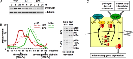
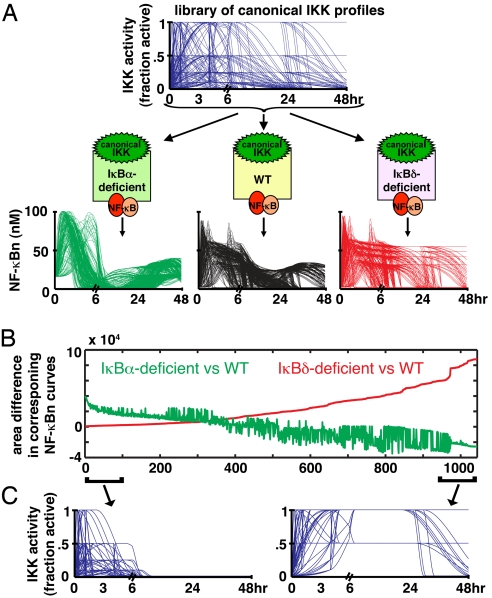
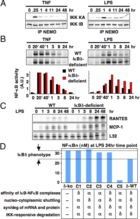
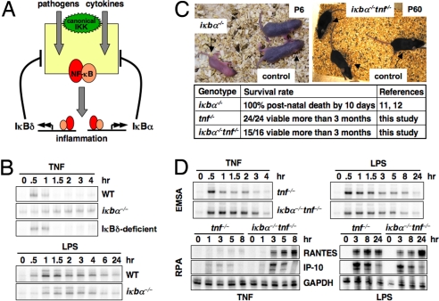
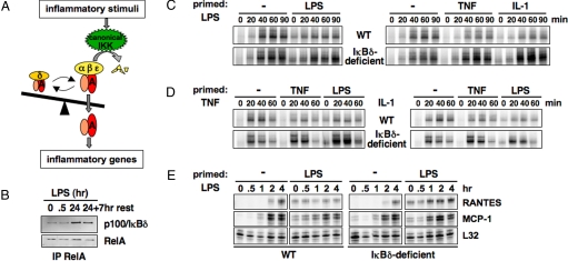
Mammalian signaling networks contain an abundance of negative feedback regulators that may have overlapping ("fail-safe") or specific functions. Within the NF-kappaB signaling module, IkappaB alpha is known as a negative feedback regulator, but the newly characterized inhibitor IkappaB delta is also inducibly expressed in response to inflammatory stimuli. To examine IkappaB delta's roles in inflammatory signaling, we mathematically modeled the 4-IkappaB-containing NF-kappaB signaling module and developed a computational phenotyping methodology of general applicability. We found that IkappaB delta, like IkappaB alpha, can provide negative feedback, but each functions stimulus-specifically. Whereas IkappaB delta attenuates persistent, pathogen-triggered signals mediated by TLRs, the more prominent IkappaB alpha does not. Instead, IkappaB alpha, which functions more rapidly, is primarily involved in determining the temporal profile of NF-kappaB signaling in response to cytokines that serve intercellular communication. Indeed, when removing the inducing cytokine stimulus by compound deficiency of the tnf gene, we found that the lethality of ikappab alpha(-/-) mouse was rescued. Finally, we found that IkappaB delta provides signaling memory owing to its long half-life; it integrates the inflammatory history of the cell to dampen NF-kappaB responsiveness during sequential stimulation events.


Conflict of interest statement

The authors declare no conflict of interest.

Figures

Fig. 1.
Fig. 1.
Characterization of IκBδ as a negative feedback regulator. (A) Western blots of whole cell extracts prepared from MEFs stimulated chronically with indicated inflammatory agents, TNF (1 ng/mL), IL-1 (1 ng/mL), LPS (100 ng/mL). Protein levels of p100/IκBδ increase whereas α-tubulin does not. (B) Chromatogram of gel-filtration studies used to separate LPS-stimulated or -unstimulated MEF cytosolic extracts. The graph shows the results from quantitated Western blots against p100 and IκBα. Thyroglobulin (670 kDa) and bovine gamma globulin (159 kDa) served as molecular size standards. (Right) A sample Western blot with indicated gel filtration fractions with antibodies against the indicated NF-κB/IκB proteins. (C) A diagram of the 4 IκB-containing NF-κB signaling module. Both pathogen-derived substances and inflammatory cytokines lead to canonical IKK-mediated NF-κB activation via the inducible degradation of 3 IκB proteins IκBα, IκBβ, and IκBε. Postinduction attenuation is mediated by an overlapping set of IκB proteins: IκBα, IκBε, and IκBδ.
Fig. 2.
Fig. 2.
Phenotyping negative feedback regulators within signaling dynamics. (A) A library of 1,044 hypothetical canonical IKK input curves (Top) over a 48-h time course (see Fig. S2A for details) was generated and fed into wild-type, IκBα-, or IκBδ-deficient computational models (Middle) to calculate the corresponding 1044 NF-κB “outputs” for each model (Bottom). (B) A plot of the quantitated NF-κB phenotype for each canonical IKK curve in IκBα or IκBδ-deficient cells, as predicted by model simulations. Differences of NF-κB activity between wild-type and knockout cells (“the phenotype”) were calculated for each canonical IKK profile and plotted after sorting the canonical IKK profiles for increasing NF-κB phenotypes in IκBδ-deficient models. (C) Time course plots of the canonical IKK activity profiles that gave rise to the least or maximum IκBδ-phenotype, as defined by the bottom and top 10% of the curves found in B.
Fig. 3.
Fig. 3.
IκBδ mediates NF-κB attenuation stimulus-specifically. (A) IKK kinase activity assays (IKK KA) from MEFs chronically stimulated with TNF (1 ng/mL) or LPS (100 ng/mL). NEMO-associated kinase complex immunoprecipitated from extracts prepared at the indicated times, and used for a kinase assay with GST-IκBα (1–54) as a substrate. Equal loading was confirmed by immunoblotting against IKK1 (IKK IB). (B) NF-κB DNA binding activities induced by chronic TNF (1 ng/mL) or LPS (100 ng/mL) stimulation in wild-type and IκBδ-deficient MEFs were monitored by EMSA. In the case of TNF, the first phase (20′, 40′ and 1-h time points) and second phase (3-, 8-, and 24-h time points) are separated by a transient trough in activity, as shown in ref. . In the case of LPS, an elongated NF-κB activity spans the entire time course. Signals were quantitated and graphed relative to resting cells. (C) RPA to monitor the expression of NF-κB-responsive inflammatory genes chronically induced by LPS (100 ng/mL) stimulation in wild-type and IκBδ-deficient MEFs. (D) Computational analyses of in silico chimeras reveal the relative importance of kinetic rate constants for IκBδ-mediated attenuation of LPS signaling. Chimeric mutants were made by swapping rate constants of IκBα to those of IκBδ as indicated. NF-κB activities induced by 24-h LPS stimulation were plotted for each chimera.
Fig. 4.
Fig. 4.
Distinct functions of IκBα and IκBδ in pathogenic and cytokine signaling. (A) IκBα and IκBδ have distinct functions in regulating NF-κB activation: We propose that IκBα is primarily involved in providing negative feedback in response to cytokine-induced signaling; IκBδ is important in mediating negative feedback upon stimulation by pathogens. (B) NF-κB DNA binding activities induced by 15-min transient TNF (1 ng/mL) or chronic LPS (100 ng/mL) stimulation in wild-type and mutant MEFs were monitored by EMSA. (C) Six-day-old iκbα−/− mice were moribund and died at 7 days. The 60-day-old iκbα−/−tnf−/− mice were indistinguishable from littermate control mice with no observable inflammation and organ abnormalities. (D) NF-κB DNA binding activities (Upper) and expression of NF-κB-responsive inflammatory genes (Lower) induced by 15-min transient TNF (1 ng/mL) or chronic LPS (100 ng/mL) stimulation in iκbα−/−tnf−/− and tnf−/− BMDMs were monitored by EMSA or RPA, respectively.
Fig. 5.
Fig. 5.
IκBδ integrates inflammatory history to limit NF-κB activation. (A) A schematic model to describe that the balance between IKK-responsive and -unresponsive RelA:p50 complexes determines the outcome of inflammatory exposure. (B) Western blots of immunoprecipitates to monitor p100/IκBδ associated with RelA during an LPS stimulation time course. RelA was immunoprecipitated from MEF whole cell extracts prepared at indicated time points. The last time point follows a 7-h rest period in endotoxin-free medium subsequent to PBS washes. (C) NF-κB activity induced by chronic LPS (100 ng/mL) in inflammation-primed wild-type and IκBδ-deficient MEFs. Cells were primed as in B. Nuclear extracts were prepared and NF-κB DNA binding activities were compared between naïve and primed cells by EMSA. (D) NF-κB activity induced by chronic TNF (1 ng/mL) and IL-1 (1 ng/mL) in inflammation-primed wild-type and IκBδ-deficient cells. Experiments were designed as in C. (E) NF-κB target gene expression monitored by RPA to assess the functional consequence of priming. Wild type and IκBδ-deficient MEFs were primed with LPS as in C, and LPS-induced (100 ng/mL) inflammatory gene expression was analyzed in naïve and primed cells at indicated time points.

References

    1. Hoffmann A, Baltimore D. Circuitry of nuclear factor kappaB signaling. Immunol Rev. 2006;210:171–186. - PubMed
    1. Basak S, et al. A fourth IkappaB protein within the NF-kappaB signaling module. Cell. 2007;128:369–381. - PMC - PubMed
    1. Savinova OV, Hoffmann A, Ghosh G. Mol Cell. 2009. The Nfkb1 and Nfkb2 proteins p105 and p100 function as the core of heterogeneous NF-kBsomes. in press. - PMC - PubMed
    1. Karin M. Nuclear factor-kappaB in cancer development and progression. Nature. 2006;441:431–436. - PubMed
    1. O'Dea EL, et al. A homeostatic model of IkappaB metabolism to control constitutive NF-kappaB activity. Mol Syst Biol. 2007;3:111. - PMC - PubMed

Publication types

LinkOut - more resources