. 2009 Jun 16;106(24):9625-30.
doi: 10.1073/pnas.0904165106. Epub 2009 Jun 1.

The structure of Aquifex aeolicus sulfide:quinone oxidoreductase, a basis to understand sulfide detoxification and respiration

Affiliations

The structure of Aquifex aeolicus sulfide:quinone oxidoreductase, a basis to understand sulfide detoxification and respiration

Marco Marcia et al. Proc Natl Acad Sci U S A. .

Abstract

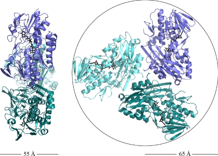
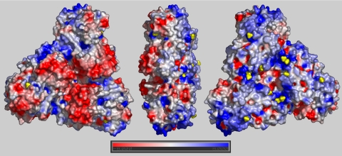
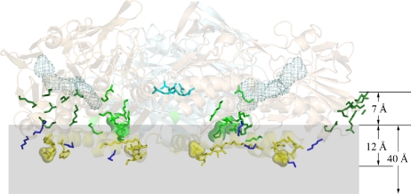
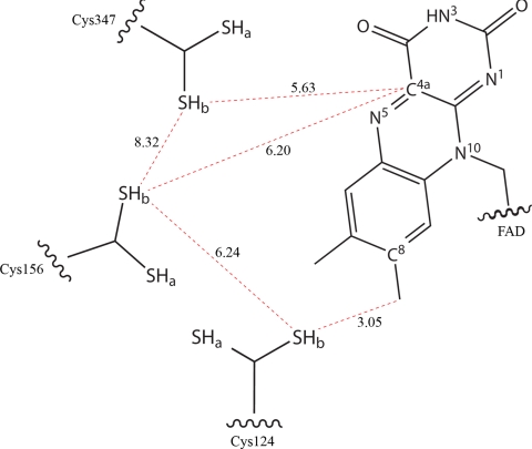
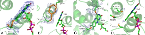
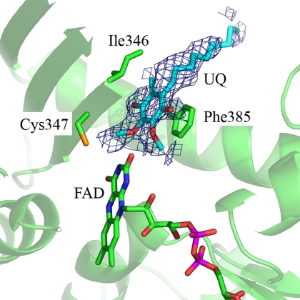
Sulfide:quinone oxidoreductase (SQR) is a flavoprotein with homologues in all domains of life except plants. It plays a physiological role both in sulfide detoxification and in energy transduction. We isolated the protein from native membranes of the hyperthermophilic bacterium Aquifex aeolicus, and we determined its X-ray structure in the "as-purified," substrate-bound, and inhibitor-bound forms at resolutions of 2.3, 2.0, and 2.9 A, respectively. The structure is composed of 2 Rossmann domains and 1 attachment domain, with an overall monomeric architecture typical of disulfide oxidoreductase flavoproteins. A. aeolicus SQR is a surprisingly trimeric, periplasmic integral monotopic membrane protein that inserts about 12 A into the lipidic bilayer through an amphipathic helix-turn-helix tripodal motif. The quinone is located in a channel that extends from the si side of the FAD to the membrane. The quinone ring is sandwiched between the conserved amino acids Phe-385 and Ile-346, and it is possibly protonated upon reduction via Glu-318 and/or neighboring water molecules. Sulfide polymerization occurs on the re side of FAD, where the invariant Cys-156 and Cys-347 appear to be covalently bound to polysulfur fragments. The structure suggests that FAD is covalently linked to the polypeptide in an unusual way, via a disulfide bridge between the 8-methyl group and Cys-124. The applicability of this disulfide bridge for transferring electrons from sulfide to FAD, 2 mechanisms for sulfide polymerization and channeling of the substrate, S(2-), and of the product, S(n), in and out of the active site are discussed.


Conflict of interest statement

The authors declare no conflict of interest.

Figures

Fig. 1.
Fig. 1.
Dimensions of the SQR trimer (cartoon representation with each monomer colored in a different shade of blue). The trimer has a thickness of ≈55 Å (Left), whereas when seen from its soluble face (Right), it can be inscribed in a circle with a radius of about 65 Å. All figures showing the structure were generated with PyMOL (www.pymol.org).
Fig. 2.
Fig. 2.
Electrostatic surface potential of the trimeric unit, calculated by the software GRASP. (Left) The view from the solvent side shows the overall negative (red) surface of the Rossmann fold domains. Rotation of the trimer by 180° (Right) shows the domain that mediates the interaction with the membrane. It has an overall intense positive charge (blue), with helices 376–395 and 400–412 more neutral (white). Sulfate ions and the Mes molecules are shown in yellow. Solvent molecules were not included in the calculation of the electrostatic potential.
Fig. 3.
Fig. 3.
Membrane-binding motifs of the SQR trimer. The membrane is indicated in gray. The overall trimer is in a cartoon semitransparent representation. The side chains of Arg-204 of all monomers are represented in cyan sticks to highlight the central trimerization contacts. Other residues and molecules are shown only for the 2 subunits in the foreground for better clarity of visualization. The FAD is in cyan mesh. The side chains of the residues and the molecules belonging to the different membrane-interacting motifs are shown as sticks. For sulfate groups a semitransparent surface is also shown. Different colors are attributed to the different structural motifs. In particular the N-terminal domain is dark green, the inner domain binding 1 sulfate ion and 1 MES molecule is light green, the 4 conserved lysines are dark blue, and the base of the trimer body is yellow. The distances were calculated from the plane of the sulfur atoms of the MES molecules, respectively, to the Cα atom of Arg-204 and the C3B atom of the maltose head of DDM (chain A). An approximate value (40 Å) indicates the membrane thickness according to White and Wimley (23).
Fig. 4.
Fig. 4.
Schematic representation of the distances (red dotted lines) between relevant Cys and FAD atoms in the catalytic site. Distances are in angstroms. The side chains of Cys-124, Cys-156, and Cys-347 are in a double conformation (-SHa/-SHb).
Fig. 5.
Fig. 5.
Electron densities in the sulfide oxidation site. The protein monomer is in a cartoon semitransparent representation in green. The FAD and the side chains of the relevant residues are in sticks, color-coded according to the atom type (C, green; N, blue; O, red; S, orange; and P, magenta). (A–C) Simulated annealing 2FoFc electron density omit maps drawn at 1.0 σ contour level in blue mesh. (A) The connection between the sulfhydryl group of Cys-124 (chain A) in one conformation and the C8M group of FAD through a putative S atom. (B) The electron density extending from the sulfhydryl group of Cys-156 (chain A) and interpreted as a covalently bound cyclooctasulfur ring. The second S atom of the chain would be trivalent, as explained in the text. (C) The sulfide oxidation site of chain D. An elongated electron density peak is prolonging the sulfhydryl group of Cys-347, whereas an only weaker density is connected to Cys-156. (D) Anomalous difference map around the catalytic site calculated from a dataset collected at 6.5 keV and shown at 3.0 σ contour level in blue mesh. Anomalous peaks are visible for S atoms of cysteines and methionines and for the phosphate groups of the FAD. One additional peak is present between the sulfhydryl group of Cys-124 and the C8M group of FAD, indicating that a relatively heavy atom with residual anomalous scattering at 6.5 keV, possibly S, is present in the structure.
Fig. 6.
Fig. 6.
The quinone-binding site (color code as in Fig. 5). The simulated annealing 2FoFc electron density omit map is shown in blue at 0.8 σ contour level, and a model for the quinone molecule is in light blue sticks.

Similar articles

Cited by

References

    1. Kimura H. Hydrogen sulfide as a neuromodulator. Mol Neurobiol. 2002;26:13–19. - PubMed
    1. Shahak Y, Hauska G. Sulfide oxidation from cyanobacteria to humans: Sulfide-quinone oxidoreductase (SQR) In: Hell R, Dahl C, Knaff DB, Leustek T, editors. Advances in Photosynthesis and Respiration. Vol 27. Heidelberg: Springer; 2008. pp. 319–335.
    1. Nübel T, Klughammer C, Huber R, Hauska G, Schütz M. Sulfide:quinone oxidoreductase in membranes of the hyperthermophilic bacterium Aquifex aeolicus (VF5) Arch Microbiol. 2000;173:233–244. - PubMed
    1. Wang R. Two's company, three's a crowd: Can H2S be the third endogenous gaseous transmitter? FASEB J. 2002;16:1792–1798. - PubMed
    1. Griesbeck C, et al. Mechanism of sulfide-quinone reductase investigated using site-directed mutagenesis and sulfur analysis. Biochemistry. 2002;41:11552–11565. - PubMed

Publication types