. 2009 Jun 16;106(24):9767-72.
doi: 10.1073/pnas.0902031106. Epub 2009 Jun 1.

Aberrant epithelial morphology and persistent epidermal growth factor receptor signaling in a mouse model of renal carcinoma

Affiliations

Aberrant epithelial morphology and persistent epidermal growth factor receptor signaling in a mouse model of renal carcinoma

Zachary S Morris et al. Proc Natl Acad Sci U S A. .

Abstract

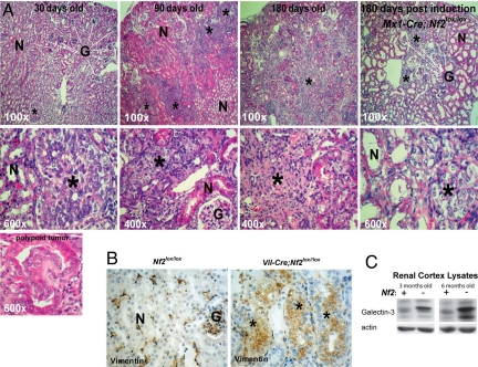
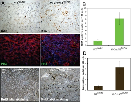
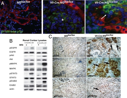
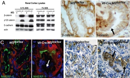
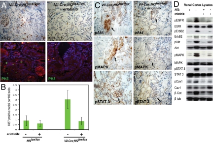
The epidermal growth factor receptor (EGFR) has frequently been implicated in hyperproliferative diseases of renal tubule epithelia. We have shown that the NF2 tumor suppressor Merlin inhibits EGFR internalization and signaling in a cell contact-dependent manner. Interestingly, despite the paucity of recurring mutations in human renal cell carcinoma (RCC), homozygous mutation of the NF2 gene is found in approximately 2% of RCC patient samples in the Sanger COSMIC database. To examine the roles of Merlin and EGFR in kidney tumorigenesis, we generated mice with a targeted deletion of Nf2 in the proximal convoluted epithelium using a Villin-Cre transgene. All of these mice developed intratubular neoplasia by 3 months, which progressed to invasive carcinoma by 6-10 months. Kidneys from these mice demonstrated marked hyperproliferation and a concomitant increase in label-retaining putative progenitor cells. Early lumen-filling lesions in this model exhibited hyperactivation of EGFR signaling, altered solubility of adherens junctions components, and loss of epithelial polarity. Renal cortical epithelial cells derived from either early or late lesions were dependent on EGF for in vitro proliferation and were arrested by pharmacologic inhibition of EGFR or re-expression of Nf2. These cells formed malignant tumors upon s.c. injection into immunocompromised mice before in vitro passage. Treatment of Vil-Cre;Nf2(lox/lox) mice with the EGFR inhibitor erlotinib halted the proliferation of tumor cells. These studies give added credence to the role of EGFR signaling and perhaps Nf2 deficiency in RCC and describe a rare and valuable mouse model for exploring the molecular basis of this disease.


Conflict of interest statement

The authors declare no conflict of interest.

Figures

Fig. 1.
Fig. 1.
Targeted loss of Nf2 in renal tubule epithelia gives rise to vimentin- and galectin-3–positive tumors that progress to invasive carcinoma. (A) All Vil-Cre;Nf2lox/lox mice developed small intratubular neoplastic growths of atypical epithelial cells by 30 days of age. These grew to large tumors within 3 months and progressed to invasive carcinomas by 6 months. Renal tumors in these mice consistently grew into the tubule lumen, often initiating with a polypoid morphology (Lower Left). The Vil-Cre;Nf2lox/lox kidney phenotype is recapitulated in inducible Mx1-Cre;Nf2lox/lox mice, which target Cre-recombinase expression to the collecting duct (Right). (B) Tumors in Vil-Cre;Nf2lox/lox kidneys stain positive by immunohistochemistry for vimentin. In Nf2lox/lox control kidneys vimentin is seen only in glomeruli, arterioles, and interstitial fibroblasts. (C) Renal cortex lysates from Vil-Cre;Nf2lox/lox mice display increased expression of galectin-3 by immunoblot. Together, vimentin and galectin-3 are histologic markers for clear cell RCC in humans (33). For A and B, the sections shown are representative of 3 mice × 2 slides per mouse × 3 sections per slide × 2 kidneys per section × ≈10 lesions per kidney = ≈360 lesions examined. N = normal tubules, G = glomeruli. *Lesion/tumor.
Fig. 2.
Fig. 2.
Vil-Cre;Nf2lox/lox kidneys display heightened epithelial cell proliferation and an increased number of label-retaining putative progenitor cells. (A) Three-month-old Vil-Cre;Nf2lox/lox kidneys displayed increased proliferation of cells mostly localized to lumen-filling neoplastic growths, as measured by nuclear staining of Ki67 (immunohistochemistry) and PH3 (green, immunofluorescence). Renal epithelial cells are displayed in red on immunofluorescence images. (B) Quantification per 100 cells revealed a ≈7-fold increase in Ki67-positive nuclei in the Vil-Cre;Nf2lox/lox renal cortex relative to controls. (C and D) BrdU pulse–chase experiments (1-week pulse, 2-week chase), previously reported to identify putative progenitor cells (57), demonstrated a ≈5-fold increase in the number of label-retaining cells in the Vil-Cre;Nf2lox/lox renal cortex relative to controls. The sections shown are representative of multiple mice, sections, and lesions as defined in the legend to Fig. 1.
Fig. 3.
Fig. 3.
Tumors in Vil-Cre;Nf2lox/lox kidneys are foci of activated EGFR signaling. (A) Immunofluorescent detection using the pY100 antibody (green) revealed a marked increase in phosphotyrosine signaling in early (Right) and late (Middle) lesions in Vil-Cre;Nf2lox/lox kidneys. Renal epithelial cells are displayed in red. (B) Immunoblot demonstrated activation of EGFR, its preferred dimerization partner ErbB2, and its downstream effectors in renal cortex lysates from 3- and 6-month-old Vil-Cre;Nf2lox/lox mice. (C) Immunohistochemical detection of phosphorylated Akt, MAPK, and STAT-3 in paraffin sections of 3-month-old kidneys demonstrated that hyperactivation of these EGFR effectors was confined to neoplastic lesions in Vil-Cre;Nf2lox/lox mice. The sections shown are representative of multiple mice, sections and lesions as defined in the legend to Fig. 1.
Fig. 4.
Fig. 4.
Tumor cells in Vil-Cre;Nf2lox/lox kidneys exhibit increased solubility of AJ components and a loss of polarity. (A) Rigorous 1% SDS solubilization demonstrated equivalent expression of AJ components. A gentler 0.1% SDS solubilization revealed stark differences in the solubility of the AJ components, p120- and β-catenin, between Vil-Cre;Nf2lox/lox and Nf2lox/lox kidney lysates. (B) Immunohistochemical staining demonstrated that this change in solubility correlated with a loss of β-catenin localization to cell–cell contacts in neoplastic cells of 3-month-old Vil-Cre;Nf2lox/lox mice. (C) Histologic examination of the apical markers NHERF-1 (green, immunofluorescence) and ezrin (immunohistochemistry) showed a loss of cell polarity in early intratubule growths arising in Vil-Cre;Nf2lox/lox kidneys. Renal epithelial cells are displayed in red on immunofluorescence images. The sections shown are representative of multiple mice, sections, and lesions as defined in the legend to Fig. 1.
Fig. 5.
Fig. 5.
Erlotinib treatment of Vil-Cre;Nf2lox/lox mice restores wild-type levels of proliferation to Nf2-deficient renal tubule epithelia. (A and B) Twelve- to 16-week-old Vil-Cre;Nf2lox/lox mice were treated by i.p. injection of 100 mg/kg erlotinib twice daily for 10 days. Erlotinib treatment restored wild-type levels of proliferation in Vil-Cre;Nf2lox/lox mice, as assayed by quantification of Ki67 (immunohistochemistry) and PH3 (green, immunofluorescence) positive nuclei per 100 cells. Statistical significance was determined by one-tailed, independent samples, parametric t test, P = 0.00085. (C) Renal tumors in erlotinib-treated mice exhibited decreased activation of EGFR signaling effectors by immunohistochemistry. The sections shown are representative of multiple mice, sections, and lesions as defined in the legend to Fig. 2. (D) Immunoblot of 0.1% SDS renal cortex lysates confirmed decreased activation of EGFR, its dimerization partner ErbB2, and its downstream effectors in erlotinib treated Vil-Cre;Nf2lox/lox mice as compared with untreated controls. Interestingly, targeting EGFR signaling also seemed to normalize the solubility of β-catenin.

Similar articles

Cited by

References

    1. Cohen HT, McGovern FJ. Renal-cell carcinoma. N Engl J Med. 2005;353:2477–2490. - PubMed
    1. Valladares Ayerbes M, et al. Origin of renal cell carcinomas. Clin Transl Oncol. 2008;10:697–712. - PubMed
    1. Iliopoulos O. Molecular biology of renal cell cancer and the identification of therapeutic targets. J Clin Oncol. 2006;24:5593–5600. - PubMed
    1. Motzer RJ, Bander NH, Nanus DM. Renal-cell carcinoma. N Engl J Med. 1996;335:865–875. - PubMed
    1. Takahashi M, Kahnoski R, Gross D, Nicol D, Teh BT. Familial adult renal neoplasia. J Med Genet. 2002;39:1–5. - PMC - PubMed

Publication types