. 2009 Jun 16;106(24):9878-83.
doi: 10.1073/pnas.0900330106. Epub 2009 Jun 1.

Proximity of excitatory and inhibitory axon terminals adjacent to pyramidal cell bodies provides a putative basis for nonsynaptic interactions

Affiliations

Proximity of excitatory and inhibitory axon terminals adjacent to pyramidal cell bodies provides a putative basis for nonsynaptic interactions

Angel Merchán-Pérez et al. Proc Natl Acad Sci U S A. .

Abstract

Although pyramidal cells are the main excitatory neurons in the cerebral cortex, it has recently been reported that they can evoke inhibitory postsynaptic currents in neighboring pyramidal neurons. These inhibitory effects were proposed to be mediated by putative axo-axonic excitatory synapses between the axon terminals of pyramidal cells and perisomatic inhibitory axon terminals [Ren M, Yoshimura Y, Takada N, Horibe S, Komatsu Y (2007) Science 316:758-761]. However, the existence of this type of axo-axonic synapse was not found using serial section electron microscopy. Instead, we observed that inhibitory axon terminals synapsing on pyramidal cell bodies were frequently apposed by terminals that established excitatory synapses with neighbouring dendrites. We propose that a spillover of glutamate from these excitatory synapses can activate the adjacent inhibitory axo-somatic terminals.


Conflict of interest statement

The authors declare no conflict of interest.

Figures
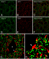
Fig. 1.
Fig. 1.
Single-plane confocal images showing immunostaining for VGluT1, GAT1, and VGAT in layer II of the mouse visual cortex. (A and D) VGluT1-ir appears in punctate structures distributed in the neuropil, as well as around unlabeled pyramidal cell somata and apical dendrites. (B and E) GAT1- and VGAT-ir are also present in puncta around cell bodies and in the neuropil. Merging of the paired images (A and B) and (D and E) shows that VGluT1 and GAT1 (C), or VGluT1 and VGAT (F), do not colocalize, even though numerous VGluT1-positive puncta are contiguous to GAT1- or VGAT-positive puncta. (G) Higher magnification of the pyramidal cell soma labeled with an asterisk in (C). The close apposition between some of the punctate structures that surround the cell body is clearly evident. The yellow, partially overlapping areas of contiguous red (GAT1) and green (VGluT1) profiles, suggest the intimate apposition of the 2 types of terminals. The arrow in (G) and (H) points to the same GAT1-positive terminal that is apposed to the cell soma and to several VGluT1-positive puncta. [Scale bar: 10 μm from (A) to (F), 3 μm in (G), and 1 μm in (H).]
Fig. 2.
Fig. 2.
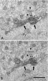
(A and B) Ultrastructural localization of VGAT in an axon terminal that makes a symmetric synapse with a pyramidal cell soma (Py). (A) and (B) are 2 consecutive ultrathin sections. The labeled terminal is filled with the dark DAB precipitate that obscures the internal details, although a mitochondrion (M), synaptic vesicles and 2 axo-somatic synapses (single arrows) can be distinguished. An unlabeled terminal (asterisks) is apposed to the labeled terminal and it makes an asymmetric (excitatory) synapse with a nearby dendritic element, probably a dendritic spine. The arrowheads point to the postsynaptic density of the asymmetric synapse. Note that at the membrane of the VGAT-ir terminal adjacent to the unlabeled terminal (double arrows) it is hard to ascertain whether a putative synaptic contact is present because the DAB reaction product is too electron dense. (Scale bar, 0.5 μm.)
Fig. 3.
Fig. 3.
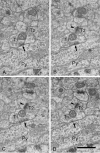
(A–D) Serial ultrathin sections showing an axo-somatic symmetric (inhibitory) synapse (arrows) and an adjacent axo-dendritic asymmetric (excitatory) synapse (arrowheads). Axon terminals forming symmetric and asymmetric synapses are indicated as T1 and T2, respectively. T2 is directly apposed to the axo-somatic bouton T1. Note that the postsynaptic density of the asymmetric synapse (arrowheads) is close to the membrane of the axo-somatic terminal (T1) and that there are no glial or neural profiles interposed. Py, pyramidal cell soma. (Scale bar, 0.5 μm.)
Fig. 4.
Fig. 4.
Three-dimensional reconstruction of an axo-somatic terminal and a neighbouring axo-dendritic synapse. (A) and (B) are 2 views at different angles (a 55° clockwise rotation through the vertical axis) to show a portion of a pyramidal cell body (gray), an axo-somatic inhibitory terminal (red), an excitatory axon terminal (green), and a spiny dendritic segment (pale brown). Note the close apposition between the axo-somatic terminal, the excitatory axon terminal, and a dendritic spine in the region indicated by arrows. (C) and (D) are paired with (A) and (B), respectively, where the axon terminals and dendritic structures have been made transparent to show the location of the postsynaptic densities (PSDs) associated with the 2 axon terminals. The PSD of the inhibitory synapse (red) is located in the pyramidal cell body, in front of the axo-somatic terminal. The PSD of the excitatory synapse (green) is located in the dendritic spine apposed to the excitatory axon terminal. This excitatory synapse is in close proximity to the inhibitory axo-somatic terminal. Notice that no axo-axonic synapse was observed between the 2 axon terminals. (Scale bar, 0.5 μm.)
Fig. 5.
Fig. 5.
Distribution of measurements derived from the 19 of the 48 axo-somatic terminals that were apposed by an excitatory terminal. (A) Comparative distribution of the shortest distances between the PSDs of asymmetric synapses and the membrane of the adjacent axo-somatic terminal (circles), and of the distances between the PSDs of asymmetric synapses and of the symmetric axo-somatic synapses (triangles). (B) Cumulative distribution of the minimum distances between the PSDs of axo-dendritic synapses and the membrane of the neighbouring axo-somatic terminals. All distances are smaller than 0.2 μm and while 72% are smaller than 0.1 μm, 56% are less than 0.05 μm. (C) Cumulative distribution of the minimum distances between the PSDs of adjacent axo-dendritic asymmetric and axo-somatic symmetric synapses. Note that the distances in (C) are 10 times that in (B).
Fig. 6.
Fig. 6.
Schematic representation of the relationship between excitatory and inhibitory synapses in the perisomatic region of a pyramidal cell. A glutamatergic terminal (Glu) establishes an excitatory synapse with a dendritic spine, which is recognized at the electron microscope level by the presence of a thick PSD (asymmetric synapse). These terminal and spine are apposed to the membrane of an axo-somatic GABAergic terminal (GABA) that establishes an inhibitory synapse with the cell body of the pyramidal cell (thinner PSD; symmetric synapse). The spillover of glutamate (green dots) from the excitatory synapse might activate the extrasynaptic receptors located on the membrane of the axo-somatic terminal. Finally, the activation of these receptors might cause the release of GABA (red dots) from the axo-somatic terminal.

Similar articles

Cited by

References

    1. DeFelipe J, Jones EG. Cajal on the Cerebral Cortex. New York: Oxford Univ Press; 1988.
    1. DeFelipe J, Fariñas I. The pyramidal neuron of the cerebral cortex: Morphological and chemical characteristics of the synaptic inputs. Prog Neurobiol. 1992;39:563–607. - PubMed
    1. Spruston N. Pyramidal neurons: Dendritic structure and synaptic integration. Nat Rev Neurosci. 2008;9:206–221. - PubMed
    1. Ren M, Yoshimura Y, Takada N, Horibe S, Komatsu Y. Specialized inhibitory synaptic actions between nearby neocortical pyramidal neurons. Science. 2007;316:758–761. - PubMed
    1. Ribak CE. Aspinous and sparsely-spinous stellate neurons in the visual cortex of rats contain glutamic acid decarboxylase. J Neurocytol. 1978;7:461–478. - PubMed

Publication types