. 2009 Jul 31;284(31):20540-7.
doi: 10.1074/jbc.M109.022541. Epub 2009 Jun 1.

Transcriptional regulation of the novel Toll-like receptor Tlr13

Affiliations

Transcriptional regulation of the novel Toll-like receptor Tlr13

Zhongcheng Shi et al. J Biol Chem. .

Abstract

Little has been known about Tlr13 (Toll-like receptor 13), a novel member of the Toll-like receptor family. To elucidate the molecular basis of murine Tlr13 gene expression, the activity of the Tlr13 gene promoter was characterized. Reporter gene analysis and electrophoretic mobility shift assays demonstrated that Tlr13 gene transcription was regulated through three cis-acting elements that interacted with the Ets2, Sp1, and PU.1 transcription factors. Furthermore, our work suggests that these transcription factors may cooperate, culminating in maximal transcription of the Tlr13 gene. In contrast, NF-kappaB appeared to act as an inhibitor of Tlr13 transcription. Overexpression of Ets2 caused a strong increase in the transcriptional activity of the Tlr13 promoter; however, overexpression of NF-kappaB p65 dramatically inhibited it. Additionally, interferon-beta is capable of acting Tlr13 transcription, but the activated signaling of lipopolysaccharide/TLR4 and peptidoglycan/TLR2 strongly inhibited the Tlr13 gene promoter. Thus, these findings reveal the mechanism of Tlr13 gene regulation, thereby providing insight into the function of Tlr13 in the immune response to pathogen.


Figures

FIGURE 1.
FIGURE 1.
Suppression of Tlr13 mRNA expression by LPS, PGN, and bacterial lysates in macrophage RAW 264.7 cells. A, regular RT-PCR was performed to analyze Tlr13 and β-actin mRNA expression in various murine cell lines, including RAW 264.7 cells, mouse embryonic fibroblasts, and NIH 3T3. B, real time PCR analysis was performed to quantitate Tlr13 expression in RAW 264.7 cells treated with 40 μl/ml bacterial lysates, 5 μg/ml PGN, 100 ng/ml LPS, or 25 μg/ml poly(I-C) for 1 or 3 h, as indicated. Staphy, Gram-positive Staphylococcus aureus K2 strain; UPEC, Gram-negative urinary pathogenic E. coli 8NU strain. The graph shows the mean ± S.D. of three independent experiments; *, p < 0.05; **, p < 0.001. Statistical analysis was performed by Student's t test.
FIGURE 2.
FIGURE 2.
Structure of murine Tlr13 gene and determination of the Tlr13 transcription start site. A, physical map of the murine Tlr13 gene and the strategy of 5′-RACE PCR amplification are shown. Tlr13 has three exons; the transcriptional start site was mapped by 5′-RACE PCR. The oligonucleotide location used for 5′-RACE PCR was indicated by an arrow, and the size of the 211-bp PCR product was determined after sequencing. Exon I in Raw264.7 cells is 17 bp longer than it is in the NCBI data base (GenBankTM number EU588988). B, 5′-RACE PCR product was resolved on a 2% agarose gel with only one specific band. The determined transcription start site is based on the specific PCR product after sequencing.
FIGURE 3.
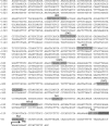
FIGURE 3.
Sequence of the 5′-flanking region of the murine Tlr13 gene. Shown is a 1.9-kb sequence of the 5′-flanking region of murine Tlr13. Underlined sequences are the potential transcription factor binding sites predicted by MatInspector software. The arrow indicates the transcription start site (TSS), which was determined by 5′-RACE.
FIGURE 4.
FIGURE 4.
Identification of essential cis-acting elements within the Tlr13 promoter. A, deletion analysis of the Tlr13 gene promoter. The truncated promoter fragments with luciferase reporter gene constructs (Luc) were contransfected with the Renilla-TK luciferase vector into RAW 264.7 cells. Firefly luciferase activity is relative to the Renilla-TK luciferase activity; values are the means ± S.D. obtained from three independent experiments. Deletions from −341 to −258 bp led to an extreme reduction of the activity. The region within −341 bp of the Tlr13 promoter contains multiple potential important transcription factor binding sites, including NF-κB, Sp1, and Est2. B, site-directed mutation analysis of the Tlr13 gene promoter. RAW 264.7 cells were transiently transfected with p65 expression vector or control vector plus the p-341 promoter plasmid or the constructs with different mutations of the Sp1, NF-κB, PU.1, or Ets2 site, respectively. Transfected cells were harvested after 24 h of transfection for the luciferase assay. Firefly luciferase activity was normalized to Renilla luciferase activity, and the values represent the means ± S.D. of three independent experiments.
FIGURE 5.
FIGURE 5.
Suppression of the Tlr13 gene promoter by LPS and PGN. A, Raw264.7 cells were transfected with Tlr13 promoter p-341 plus Renilla-TK luciferase vector by Lipofectamine 2000. Twenty-four hours after transfection, cells were treated with medium alone, 5 μg/ml PGN, 100 ng/ml LPS, 25 μg/ml poly(I-C), or 100 units/ml IFN-β. Firefly luciferase activity was assayed 6 h after treatment and normalized to Renilla luciferase activity. Data present the mean ± S.D. of three independent experiments. Statistical analysis was performed by Student's t test; *, p < 0.05; **, p < 0.005. B, all of the procedure was performed in the same way as in A except for the pretreatment with PS1145 (10 μm for 3 h) or PBS before LPS stimulation. C, Raw264.7 cells were pretreated with PS1145 (10 μm for 3 h) or left untreated (UN) and then stimulated with LPS (100 ng/ml) for 0, 1, or 3 h. The endogenous Tlr13 expression was analyzed by real time PCR.
FIGURE 6.
FIGURE 6.
Binding of PU.1 and Ets2 to the promoter. Electrophoretic mobility shift assay (EMSA) targets the identified elements with potential important for transcriptional regulation in the Tlr13 promoter, including Ets2 (A and C) and PU.1 (B). A radiolabeled double-stranded DNA probe containing the Ets2 (A and C) and PU.1 (B) binding region in the Tlr13 promoter was incubated with nuclear extracts from RAW 264.7 cells and separated on a 6% polyacrylamide gel. Specificity was determined by the addition of antibodies for supershift, a 100-fold molar excess of unlabeled cold probe, mutant probe, or other control probe as specific and nonspecific competitors, as indicated above the corresponding lanes. The arrowheads indicate specific complexes with the specific element. Control, probe only without nuclear extracts. C, overexpression of NF-κB p65 is capable of inhibiting Ets2 binding in a dose-dependent manner. RAW264.7 cells were transfected with empty vector and p65 at a ratio of 99:1, 90:10, and 0:100 for lanes 3–5, respectively. NS, nonspecific.
FIGURE 7.
FIGURE 7.
Characterization of the transcription factors in the murine Tlr13 gene promoter. A, RAW 264.7 cells were transfected with Tlr13 promoter p-341 plus expression vector for Ets2, p65, PU.1 and Sp1, respectively, and with different combinations of expression vectors. Transfected cells were harvested after 24 h for the luciferase assay. B, Raw264.7 cells were treated with LPS (100 ng/ml) for 0.5 h. Cells were lysed at 48 h post-transfection. Total protein was harvested and was used to perform immunoblotting (IB) and immunoprecipitation (IP) by the antibodies as indicated. Similar results were obtained in three independent experiments. C, RAW 264.7 cells were transfected with wild type p-341 or Ets2 site mutation p-341 plasmids plus different doses of Ets2 expression construct. D, RAW 264.7 cells were transfected with wild type p-341 or Ets2 site mutation p-341 plasmids plus expression vectors for Ets2, Raf-1, and the combination of Ets2 and Raf-1. Transfected cells were harvested after 24 h for the luciferase assay.
FIGURE 8.
FIGURE 8.
A model of Tlr13 transcriptional regulation. Tlr13 gene transcription is regulated through three cis-acting elements that interact with the Ets2, Sp1, and PU.1 transcription factors. In contrast, NF-κB appears to act as an inhibitor of Tlr13 transcription. The activated signaling of LPS/TLR4 and PGN/TLR2 strongly inhibit the Tlr13 gene promoter activity, perhaps through NF-κB activation. The inhibition is most likely through p65, which directly interacts with Ets2.

Similar articles

Cited by

References

    1. Janeway C. A., Jr., Medzhitov R. (2002) Annu. Rev. Immunol. 20, 197–216 - PubMed
    1. Aderem A., Ulevitch R. J. (2000) Nature 406, 782–787 - PubMed
    1. Akira S., Uematsu S., Takeuchi O. (2006) Cell 124, 783–801 - PubMed
    1. Beutler B. (2002) Curr. Opin. Hematol. 9, 2–10 - PubMed
    1. Underhill D. M., Ozinsky A. (2002) Curr. Opin. Immunol. 14, 103–110 - PubMed

Publication types

MeSH terms

LinkOut - more resources