Review
. 2009 Jun;73(2):249-99.
doi: 10.1128/MMBR.00035-08.

Ecological genomics of marine picocyanobacteria

Affiliations
Review

Ecological genomics of marine picocyanobacteria

D J Scanlan et al. Microbiol Mol Biol Rev. 2009 Jun.

Abstract

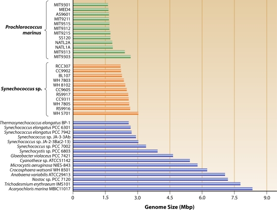
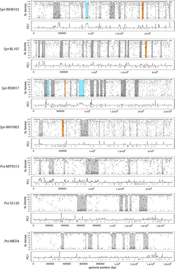
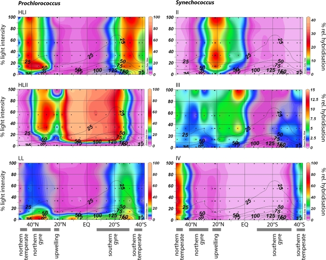
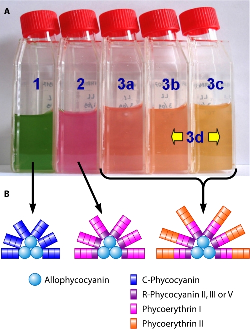
Marine picocyanobacteria of the genera Prochlorococcus and Synechococcus numerically dominate the picophytoplankton of the world ocean, making a key contribution to global primary production. Prochlorococcus was isolated around 20 years ago and is probably the most abundant photosynthetic organism on Earth. The genus comprises specific ecotypes which are phylogenetically distinct and differ markedly in their photophysiology, allowing growth over a broad range of light and nutrient conditions within the 45 degrees N to 40 degrees S latitudinal belt that they occupy. Synechococcus and Prochlorococcus are closely related, together forming a discrete picophytoplankton clade, but are distinguishable by their possession of dissimilar light-harvesting apparatuses and differences in cell size and elemental composition. Synechococcus strains have a ubiquitous oceanic distribution compared to that of Prochlorococcus strains and are characterized by phylogenetically discrete lineages with a wide range of pigmentation. In this review, we put our current knowledge of marine picocyanobacterial genomics into an environmental context and present previously unpublished genomic information arising from extensive genomic comparisons in order to provide insights into the adaptations of these marine microbes to their environment and how they are reflected at the genomic level.
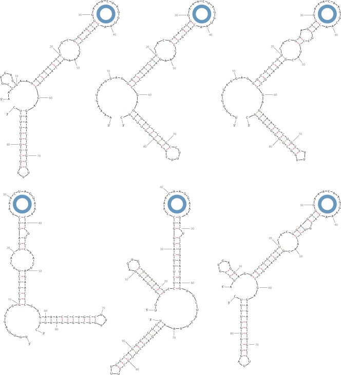

Figures

FIG. 1.
FIG. 1.
Neighbor-joining tree, based on 16S rRNA gene sequences, indicating the phylogenetic relationships among marine picocyanobacteria that are the subject of this review. Bootstrap values of >70% are shown. Strains with sequenced genomes are in boldface.
FIG. 2.
FIG. 2.
Genome sizes of marine picocyanobacteria compared to a representative selection of other cyanobacteria.
FIG. 3.
FIG. 3.
Genome plot of recently acquired genomic islands in marine Synechococcus and Prochlorococcus strains. For each genome predicted islands are highlighted in gray, except for the phycobilisome gene cluster, which is highlighted in orange. The frequency with which an ortholog appears among the 14 genomes (11 Synechococcus and three Prochlorococcus) of the curated Cyanorak database (http://www.sb-roscoff.fr/Phyto/cyanorak/) is represented by a black bar (a core gene is present in 14 genomes). Giant ORFs are highlighted in blue. Tetranucleotide frequency in overlapping 5,000 bp intervals was transformed by principal-component analysis, and the deviation in tetranucleotide frequency is plotted in the box below each genome as the first principal component (PC1). The positions of tRNA genes (purple bars) and mobility genes, such as those encoding phage integrases and transposases, are also indicated (green bars). While for marine Synechococcus strains and Prochlorococcus sp. strain MIT9313 the deviation in tetranucleotide frequency correlates well with genomic regions bounded by mobility genes and containing unique genes or orthologs common to few genomes, for the streamlined, rapidly evolving A+T-rich genomes of Prochlorococcus there is a poor correlation between tetranucleotide frequency and predicted islands. (Modified from reference with permission of the publisher.)
FIG. 4.
FIG. 4.
Dot blot hybridization data showing the distribution of HLI, HLII, and LL Prochlorococcus ecotypes and Synechococcus clades II to IV in the euphotic zone along an Atlantic Meridional cruise track (AMT 15). Contour plots indicate the percent relative hybridization. For each panel the y axis is the light intensity (percent surface irradiance) and the x axis the latitude (degrees north or south of the equator). Black contour lines indicate the depth in meters, and black dots represent sampling points. (Adapted from reference with permission of Wiley-Blackwell.)
FIG. 5.
FIG. 5.
Synechococcus pigment types and associated phycobilisome structures. (A) Photograph of cultures representative of the three main pigment types (1 to 3) and subtypes (3a to 3c). Pigment type 3d corresponds to type IV chromatic adapters that modify their pigmentation from subtype 3b (in green light) to 3c (in blue light). (B) Models of the phycobilisome structures of the three main pigment types. (Adapted from reference with permission of the publisher.)
FIG. 6.
FIG. 6.
Neighbor-joining tree indicating the phylogenetic relatedness of the different peroxiredoxins found in marine picocyanobacteria. The Cyanorak cluster numbers are indicated for each peroxiredoxin type. Color coding: green, Prochlorococcus; orange, marine Synechococcus; blue, other cyanobacteria.
FIG. 7.
FIG. 7.
Phylogenetic analysis of PII proteins in unicellular marine (Synechococcus and Prochlorococcus) and unicellular freshwater (Synechocystis sp. strain PCC6803 and Synechococcus elongatus PCC7942) cyanobacteria. The two PII proteins of E. coli were chosen as outgroups. Bootstrap values are given at nodes if at least 60. The two paralogs of Synechococcus sp. strain WH5701 are indicated in boldface. Color coding: green, Prochlorococcus; orange, marine Synechococcus; blue, other cyanobacteria; black, E. coli.
FIG. 8.
FIG. 8.
Multiple sequence alignment of cyanobacterial PII of marine Synechococcus strains (Syn), selected Prochlorococcus strains (Pro), and the two freshwater strains Synechocystis sp. strain PCC6803 and Synechococcus elongatus PCC7942 and of the two E. coli PII proteins. Sites that belong to a previously suggested cyanobacterial signature (216) are indicated by arrows. #, the functionally relevant residue Arg45, required for interaction with NAGK; $, Ser49, which becomes phosphorylated in S. elongatus PCC 7942 (78). Asterisks and numbers above the sequences mark every 10th position.
FIG. 9.
FIG. 9.
Partial alignment of a genomic fragment containing the gene clusters for assimilation pathways of major N sources for marine Synechococcus strains organized according to phylogenetic clusters (56), i.e., urea (blue), nitrate (green), nitrite (pink), and cyanate (yellow), in addition to N metabolism genes (purple), other nonrelated genes (white), and fully conserved genes flanking this region (red). Genes encoding the enzyme central to each pathway are in dark color, while genes involved in accessory functions (acquisition/cofactor biosynthesis) are in light color. Dotted lines and block arrows indicate mobile fragments of 10 to 25 genes in size that are subject to deletion/insertion events in different lineages. The upper panel presents the relevant genomic regions in Synechococcus sp. strain WH5701.
FIG. 10.
FIG. 10.
Neighbor-joining tree based on the amino acid sequences of PstS and SphX from marine and freshwater cyanobacteria. Three families of phosphate-binding proteins are evident in marine Synechococcus (PstSI, PstSII, and SphX), which suggests that these proteins are functionally distinct (e.g., they may display different affinities for the same substrate). The PstS sequences of HL and LL Prochlorococcus strains, freshwater cyanobacteria, and marine Synechococcus strains form separate clusters, while the SphX sequences all cluster together. The tree was constructed from an alignment using Clustal X v1.83. The bootstrap values were obtained through 1,000 repetitions. The tree was rooted on the PstS sequence of E. coli K-12. Sequences were extracted from BLAST searches of GenBank (www.ncbi.nlm.nih.gov/BLAST) or using Cyanorak (http://www.sb-roscoff.fr/Phyto/cyanorak/) or Cyanobase (www.kazusa.or.jp/cyanobase/). The scale bar represents 10 substitutions per 100 nucleotides. Color coding: green, Prochlorococcus; orange, marine Synechococcus; blue, other cyanobacteria; black, cyanophage sequences.
FIG. 11.
FIG. 11.
Schematic representation of the different domain structures of phosphatases present in marine and freshwater cyanobacterial genomes. At least five different families of phosphatases are represented. Notable features include the variable domain structure of the large PhoA-type phosphatases which contain UshA and 5′ nucleotidase domains. SynWH7803_0111 includes an N-terminal insertion of a phytase-like domain (COG4222), homologous to SYNW0762, which is also represented in MED4_ 0708 and MIT9312_0720. COG3391 is a potential Zn-binding domain, which suggests a requirement for Zn as a cofactor. PhoX (332) and PhoD (64) homologs are also represented in a subset of strains. The approximate molecular mass is indicated by the scale at the bottom. Color coding of sequence names: green, Prochlorococcus; orange, marine Synechococcus; blue, other cyanobacteria; black, other bacteria. a, this ORF contains a potential frameshift; b, this ORF is truncated and hence is a probable pseudogene; c, this ORF spans only the N-terminal portion of PhoX; d, this ORF is truncated at its N terminus. (Adapted from reference with permission of the publisher.)
FIG. 12.
FIG. 12.
Multiple-sequence alignment, using ClustalX v1.83, of the N-terminal region of the PhoR histidine kinase in marine picocyanobacteria. Differences in the N-terminal sequences suggest alternative subcellular locations for PhoR in different strains of picocyanobacteria. Shown at the bottom is the probability of a transmembrane domain for the first 100 amino acids (aa) of the N-terminal regions (predicted by the TMHMM server) for the three groups of organisms defined in the text.
FIG. 13.
FIG. 13.
Phylogenetic relationships among CRP family regulators of marine picocyanobacteria. The identified CRP regulators form four distinct clusters, i.e., NtcA, cluster 2049, cluster 1390, and PtrA, with decreasing levels of conservation and a further diverse group mostly composed of cluster 2546. Color coding: green, Prochlorococcus; orange, marine Synechococcus.
FIG. 14.
FIG. 14.
Marine picocyanobacterial genomic regions centered on a potential Fe regulator of the CRP family (cluster 1390). The genomic region for Synechococcus sp. strain CC9605 is essentially the same as for CC9902/BL107 except that petF, encoding ferredoxin, is located downstream of ferritin and upstream of the hypothetical proteins. For Synechococcus at least, these genes are located in a genomic island (WH7805-ISL3, BL107-ISL6, CC9605-ISL12, CC9902-ISL-11, RS9916 ISL4). futA, ABC-type iron transporter, periplasmic iron-binding protein; futB, ABC-type iron transporter, ATP-binding component; futC, ABC-type iron transporter, membrane component; feoB, ferrous iron uptake protein; isiB, flavodoxin; som, porin; trxB, thioredoxin reductase; A, putative Fe-regulated hydroxylase; B, ABC molybdenum transporter-binding component; C, HupE hydrogenase component; chp, conserved hypothetical protein.
FIG. 15.
FIG. 15.
Phylogenetic relationships among marine picocyanobacterial sigma factors. A total of 136 sigma factors from 22 strains of marine picocyanobacteria (11 Synechococcus [Syn] and 11 Prochlorococcus [Pro] strains) were analyzed. The vegetative major sigma factor SigA and the four alternative type 2 sigma factors from Synechocystis sp. strain PCC6803 were included for orientation. Clades A to E refer to different classes of closely related sigma factors from picocyanobacteria. Bootstrap numbers are given for major nodes. Color coding: green, Prochlorococcus; orange, marine Synechococcus; blue, other cyanobacteria. (See Fig. 16 for more information on clade D.)
FIG. 16.
FIG. 16.
Phylogenetic relationships among clade D (Fig. 15) type 2 alternative sigma factors. All Synechococcus factors are indicated in boldface. For easier identification, GenBank tags are given and the respective part indicating the strain is underlined. The genes encoding these proteins are probably rapidly evolving, since a series of independent duplications (black dots) in the progenitors of single strains or two strains, respectively, is suggested by similarity and phylogenetic analysis. The proteins BL107_06099 and SynCC9902_0139 differ only by a single conservative exchange (identity at DNA level, 87%). Bootstrap numbers are given for all nodes. Color coding: green, Prochlorococcus; orange, marine Synechococcus.
FIG. 17.
FIG. 17.
DNA integration into the tmRNA gene of Synechococcus sp. strain WH8102. The start of genomic island 1 is indicated, and the positions of three phage integrase genes are given by the blue boxes, together with their sequence ID. The order of ssrA acceptor (tRNA-like) and coding (mRNA-like) segments is reversed in picocyanobacteria compared to most other bacteria (94). The genetic element Synw12X (325) is integrated into the 3′ end of ssrA, far from the tRNA-like segment. Within the triplicated 33-bp attB sequences resides an inverted repeat able to fold into a secondary structure (arrows). These elements exhibit compensatory base pair changes, indicating the functional importance of the stem-loop formed by this repeat.
FIG. 18.
FIG. 18.
Predicted secondary structures of the six selected ncRNAs from Synechococcus sp. strain CC9311 belonging to the Yfr2 to -5 class. A characteristic motif is the conserved sequence 5′-GAAAC(U/A)AGG(C/U/A)AA-3′ in the single-stranded loop region labeled by the doughnut. This motif is similarly conserved between all members of this ncRNA class throughout the cyanobacterial radiation. The structures were drawn with mfold (339).

References

    1. Reference deleted.
    1. Ahlgren, N. A., and G. Rocap. 2006. Culture isolation and culture-independent clone libraries reveal new marine Synechococcus ecotypes with distinctive light and N physiologies. Appl. Environ. Microbiol. 727193-7204. - PMC - PubMed
    1. Ahlgren, N. A., G. Rocap, and S. W. Chisholm. 2006. Measurement of Prochlorococcus ecotypes using real-time polymerase chain reaction reveals different abundances of genotypes with similar light physiologies. Environ. Microbiol. 8441-454. - PubMed
    1. Aiba, H., and T. Mizuno. 1994. A novel gene whose expression is regulated by the response regulator, SphR, in response to phosphate limitation in Synechococcus species PCC7942. Mol. Microbiol. 1325-34. - PubMed
    1. Al-Taweel, K., T. Iwaki, Y. Yabuta, S. Shigeoka, N. Murata, and A. Wadano. 2007. A bacterial transgene for catalase protects translation of D1 protein during exposure of salt-stressed tobacco leaves to strong light. Plant Physiol. 145258-265. - PMC - PubMed

Publication types

LinkOut - more resources