

Challenging a paradigm: the role of DNA homology in tyrosine recombinase reactions
- PMID: 19487729
- PMCID: PMC2698419
- DOI: 10.1128/MMBR.00038-08
Challenging a paradigm: the role of DNA homology in tyrosine recombinase reactions
Abstract
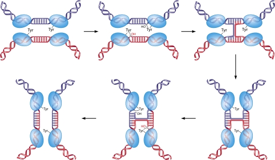
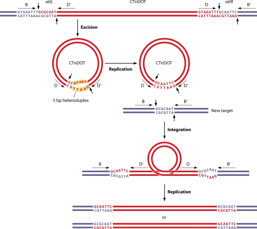
A classical feature of the tyrosine recombinase family of proteins catalyzing site-specific recombination, as exemplified by the phage lambda integrase and the Cre and Flp recombinases, is the ability to recombine substrates sharing very limited DNA sequence identity. Decades of research have established the importance of this short stretch of identity within the core regions of the substrates. Since then, several new enzymes that challenge this paradigm have been discovered and require the role of sequence identity in site-specific recombination to be reconsidered. The integrases of the conjugative transposons such as Tn916, Tn1545, and CTnDOT recombine substrates with heterologous core sequences. The integrase of the mobilizable transposon NBU1 performs recombination more efficiently with certain core mismatches. The integration of CTX phage and capture of gene cassettes by integrons also occur by altered mechanisms. In these systems, recombination occurs between mismatched sequences by a single strand exchange. In this review, we discuss literature that led to the formulation of the current strand-swapping isomerization model for tyrosine recombinases. The review then focuses on recent developments on the recombinases that challenged the paradigm that was derived from the studies of early systems.
Figures


Similar articles
-
Resolution of Mismatched Overlap Holliday Junction Intermediates by the Tyrosine Recombinase IntDOT.J Bacteriol. 2017 Apr 25;199(10):e00873-16. doi: 10.1128/JB.00873-16. Print 2017 May 15. J Bacteriol. 2017. PMID: 28242723 Free PMC article.
-
CTnDOT integrase performs ordered homology-dependent and homology-independent strand exchanges.Nucleic Acids Res. 2007;35(17):5861-73. doi: 10.1093/nar/gkm637. Epub 2007 Aug 24. Nucleic Acids Res. 2007. PMID: 17720706 Free PMC article.
-
Specific DNA cleavage mediated by the integrase of conjugative transposon Tn916.J Bacteriol. 1997 Feb;179(4):1117-25. doi: 10.1128/jb.179.4.1117-1125.1997. J Bacteriol. 1997. PMID: 9023193 Free PMC article.
-
Conjugative transposition.Annu Rev Microbiol. 1995;49:367-97. doi: 10.1146/annurev.mi.49.100195.002055. Annu Rev Microbiol. 1995. PMID: 8561465 Review.
-
A structural view of cre-loxp site-specific recombination.Annu Rev Biophys Biomol Struct. 2001;30:87-104. doi: 10.1146/annurev.biophys.30.1.87. Annu Rev Biophys Biomol Struct. 2001. PMID: 11340053 Review.
Cited by
-
Strain-specific genes of Helicobacter pylori: genome evolution driven by a novel type IV secretion system and genomic island transfer.Nucleic Acids Res. 2010 Oct;38(18):6089-101. doi: 10.1093/nar/gkq378. Epub 2010 May 16. Nucleic Acids Res. 2010. PMID: 20478826 Free PMC article.
-
Mobilization of retrotransposons as a cause of chromosomal diversification and rapid speciation: the case for the Antarctic teleost genus Trematomus.BMC Genomics. 2018 May 9;19(1):339. doi: 10.1186/s12864-018-4714-x. BMC Genomics. 2018. PMID: 29739320 Free PMC article.
-
Conservative site-specific and single-copy transgenesis in human LINE-1 elements.Nucleic Acids Res. 2016 Apr 7;44(6):e55. doi: 10.1093/nar/gkv1345. Epub 2015 Dec 15. Nucleic Acids Res. 2016. PMID: 26673710 Free PMC article.
-
DNA Transposition at Work.Chem Rev. 2016 Oct 26;116(20):12758-12784. doi: 10.1021/acs.chemrev.6b00003. Epub 2016 May 17. Chem Rev. 2016. PMID: 27187082 Free PMC article. Review.
-
Roles of Exc protein and DNA homology in the CTnDOT excision reaction.J Bacteriol. 2012 Jul;194(13):3368-76. doi: 10.1128/JB.00359-12. Epub 2012 Apr 13. J Bacteriol. 2012. PMID: 22505687 Free PMC article.
References
-
- Amin, A. A., L. G. Beatty, and P. D. Sadowski. 1990. Synaptic intermediates promoted by the FLP recombinase. J. Mol. Biol. 21455-72. - PubMed
Publication types
MeSH terms
Substances
Grants and funding
LinkOut - more resources
Full Text Sources
Other Literature Sources
