. 2009 Jul;119(7):1910-20.
doi: 10.1172/JCI35412.

Type I IFNs mediate development of postinfluenza bacterial pneumonia in mice

Affiliations

Type I IFNs mediate development of postinfluenza bacterial pneumonia in mice

Arash Shahangian et al. J Clin Invest. 2009 Jul.

Abstract

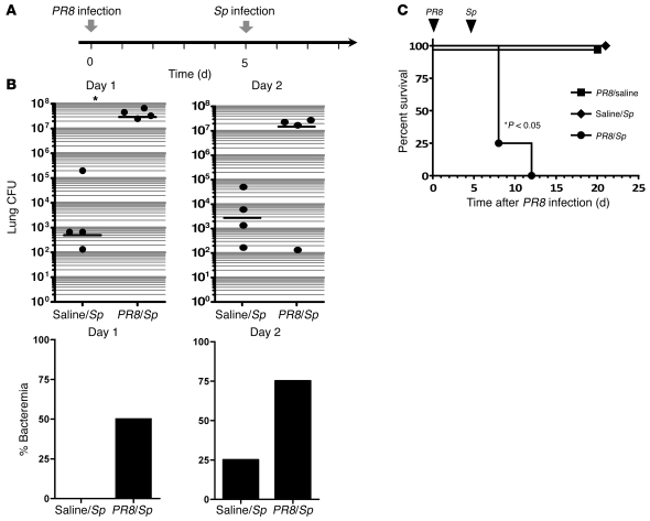
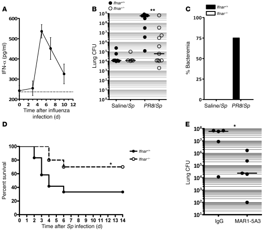
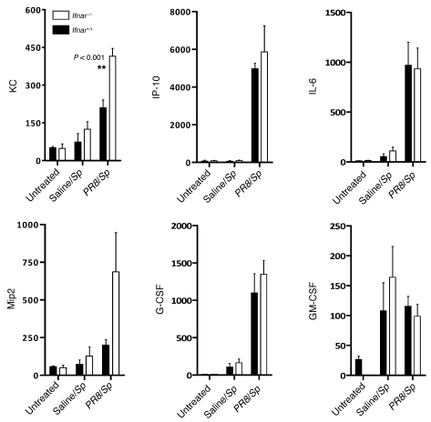
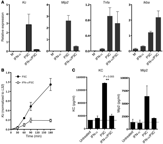
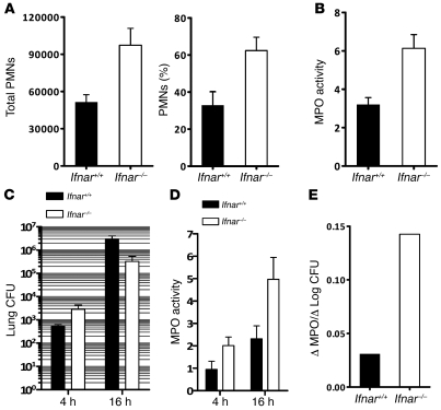
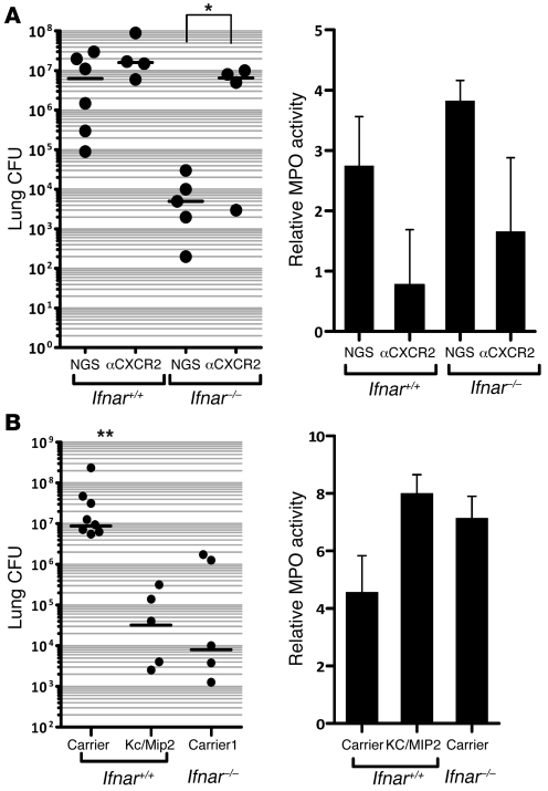
Influenza-related complications continue to be a major cause of mortality worldwide. Due to unclear mechanisms, a substantial number of influenza-related deaths result from bacterial superinfections, particularly secondary pneumococcal pneumonia. Here, we report what we believe to be a novel mechanism by which influenza-induced type I IFNs sensitize hosts to secondary bacterial infections. Influenza-infected mice deficient for type I IFN-alpha/beta receptor signaling (Ifnar-/- mice) had improved survival and clearance of secondary Streptococcus pneumoniae infection from the lungs and blood, as compared with similarly infected wild-type animals. The less effective response in wild-type mice seemed to be attributable to impaired production of neutrophil chemoattractants KC (also known as Cxcl1) and Mip2 (also known as Cxcl2) following secondary challenge with S. pneumoniae. This resulted in inadequate neutrophil responses during the early phase of host defense against secondary bacterial infection. Indeed, influenza-infected wild-type mice cleared secondary pneumococcal pneumonia after pulmonary administration of exogenous KC and Mip2, whereas neutralization of Cxcr2, the common receptor for KC and Mip2, reversed the protective phenotype observed in Ifnar-/- mice. These data may underscore the importance of the type I IFN inhibitory pathway on CXC chemokine production. Collectively, these findings highlight what we believe to be a novel mechanism by which the antiviral response to influenza sensitizes hosts to secondary bacterial pneumonia.


Figures

Figure 1
Figure 1. Primary influenza pneumonia enhances sensitivity to secondary pneumococcal infection.
(A) Experimental setup for combinatorial infection model of wild-type C57BL/6 animals. Mice were administered i.t. influenza (PR8 strain, 200 PFU) or saline, followed 5 days later by i.t. S. pneumoniae (Sp) or saline. (B) Pulmonary bacterial burden and incidence of bacteremia were measured in saline/S. pneumoniae– and PR8/S. pneumoniae–infected animals on days 1 and 2 after secondary bacterial challenge (2,000 CFU). (C) Survival was examined for 3 weeks after the initial i.t. challenge for the following 3 groups: PR8/saline, saline/S. pneumoniae, and PR8/S. pneumoniae. n = 4–8 animals per group. Data are representative of 2 independent experiments.
Figure 2
Figure 2. Influenza-infected Ifnar–/ mice are resistant to secondary pneumococcal pneumonia.
(A) Kinetics of IFN-α induction after influenza following i.t. PR8. Ifnar+/+ C57BL/6 mice were administered i.t. PR8 on day 0, and lungs were harvested at the designated time points for assessment of IFN-α levels by ELISA in lung homogenates. (B and C) Clearance of S. pneumoniae by influenza-infected Ifnar+/+ and Ifnar–/– animals. Age- and sex-matched animals of both genotypes were administered i.t. PR8 or saline, followed 5 days later with i.t. S. pneumoniae (2,000 CFU). Lungs (B) and blood (C) were harvested on day 2 following i.t. S. pneumoniae infection, for assessment of CFU. n = 8–11 animals per group (B) and n = 4 animals per group (C). **P < 0.01. Data are representative of 3 independent experiments. (D) Following i.t. PR8 and S. pneumoniae administration 5 days apart, survival was assessed for 14 days following S. pneumoniae infection. Survival rates were 70% for Ifnar–/– mice compared with 33% for Ifnar+/+ mice at 14 days. *P = 0.05, log-rank test. n = 10–12 mice per group. Data were combined from 2 separate experiments. (E) Lung CFU in animals following IFNAR neutralization of wild-type animals with MAR1-5A3. P < 0.05, 1-tailed Mann-Whitney U test. The horizontal lines represent the statistical medians.
Figure 3
Figure 3. Lung homogenates from PR8/S. pneumoniae–infected Ifnar–/– mice contain higher levels of KC and Mip2.
Production of KC and Mip2 in response to secondary pneumococcal infection, as determined by ELISA analysis of Ifnar+/+ and Ifnar–/– mice with prior influenza infection. Levels of other inflammatory cytokines were determined by multiplex cytokine protein analysis. Lung homogenates were obtained and analyzed 16 hours after secondary challenge with S. pneumoniae. n ≥ 4 mice per group.
Figure 4
Figure 4. Type I IFNs inhibit production of Kc and Mip2 but not Tnfa or Ikba transcript in P3C-stimulated BMMs.
(A) Pretreatment of BMMs with IFN-α for 30 minutes, followed by stimulation with the TLR2 ligand P3C. Kc, Mip2, Tnfa, and Ikba expression was assessed quantitative PCR 4 hours after P3C stimulation. Data are representative of 4 independently performed experiments. M, media. (B) BMMs were exposed to recombinant IFN-α for 30 minutes, followed by P3C stimulation. Levels of KC expression were examined by quantitative PCR at various time points following P3C stimulation. All values were normalized to L32 expression levels. (C) Production of KC and Mip2 protein levels in cell culture supernatants were assessed by ELISA. BMMs were exposed to IFN-α, P3C, or IFN-α and P3C for 24 hours prior to collection of supernatants for analysis.
Figure 5
Figure 5. Ifnar–/– mice have higher numbers of airspace PMNs following i.t. PR8 and S. pneumoniae.
(A) Total number and percentage of infiltrating PMNs in doubly infected Ifnar+/+ and Ifnar–/– animals were analyzed in BAL 14 hours after secondary pneumococcal challenge. (B) MPO activity was assessed in lung homogenates of doubly infected Ifnar–/– and Ifnar+/+ mice 24 hours after secondary challenge with S. pneumoniae (P < 0.05). n = 3. Data are representative of at least 2 independently performed experiments. (C) Lung homogenate CFU of doubly infected Ifnar+/+ and Ifnar–/– animals at 4 and 16 hours following S. pneumoniae challenge. (D) MPO activity of lung homogenates from doubly infected Ifnar+/+ and Ifnar–/– animals were determined at 4 and 16 hours after secondary S. pneumoniae challenge. (E) Average change in MPO activity (16 hours – 4 hours) divided by the average change in Log CFU (16 hours – 4 hours).
Figure 6
Figure 6. KC and Mip2 play a critical role in the clearance of secondary bacterial infection.
(A) Ifnar+/+ and Ifnar–/– animals were administered i.t. PR8, followed 5 days later by i.t. S. pneumoniae. Anti-Cxcr2 antibody or normal goat serum were administered i.p. 24 hours prior to i.t. S. pneumoniae challenge. Left: Lung homogenate CFU was assessed 24 hours after secondary S. pneumoniae (*P < 0.05, n = 4–6 animals per group). Right: MPO activity of lung homogenates was assessed 24 hours after S. pneumoniae infection. NGS, normal goat serum. (B) Animals were administered i.t. PR8, followed 5 days later by i.t. S. pneumoniae. Recombinant murine KC and Mip2 were given i.t. at the time of pneumococcal administration. At 24 hours following secondary S. pneumoniae challenge, lungs were collected for homogenization and determination of CFU (left; **P < 0.01; n = 5–8 mice per group) and MPO activity (right). Data are representative of 2 independent experiments. Carrier, saline carrier. Horizontal bars in the left panels represent statistical medians.

References

    1. Hamilton B.E., et al. Annual summary of vital statistics: 2005. Pediatrics. 2007;119:345–360. doi: 10.1542/peds.2006-3226. - DOI - PubMed
    1. LeVine A.M., Koeningsknecht V., Stark J.M. Decreased pulmonary clearance of S. pneumoniae following influenza A infection in mice. J. Virol. Methods. 2001;94:173–186. doi: 10.1016/S0166-0934(01)00287-7. - DOI - PubMed
    1. Sethi S. Bacterial pneumonia. Managing a deadly complication of influenza in older adults with comorbid disease. Geriatrics. 2002;57:56–61. - PubMed
    1. McCullers J.A. Insights into the interaction between influenza virus and pneumococcus. Clin. Microbiol. Rev. 2006;19:571–582. doi: 10.1128/CMR.00058-05. - DOI - PMC - PubMed
    1. Broug-Holub E., et al. Alveolar macrophages are required for protective pulmonary defenses in murine Klebsiella pneumonia: elimination of alveolar macrophages increases neutrophil recruitment but decreases bacterial clearance and survival. Infect. Immun. 1997;65:1139–1146. - PMC - PubMed

Publication types