. 2009 Jul;119(7):1974-85.
doi: 10.1172/JCI33816.

Molecular disruption of RAD50 sensitizes human tumor cells to cisplatin-based chemotherapy

Affiliations

Molecular disruption of RAD50 sensitizes human tumor cells to cisplatin-based chemotherapy

Waleed M Abuzeid et al. J Clin Invest. 2009 Jul.

Erratum in

  • J Clin Invest. 2012 Nov;122(11):4300

Abstract

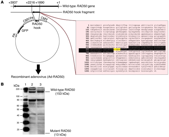
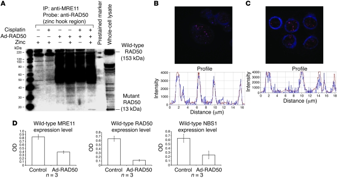
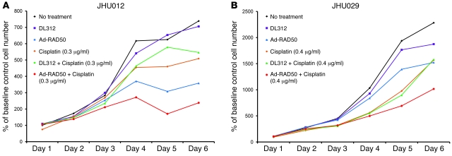
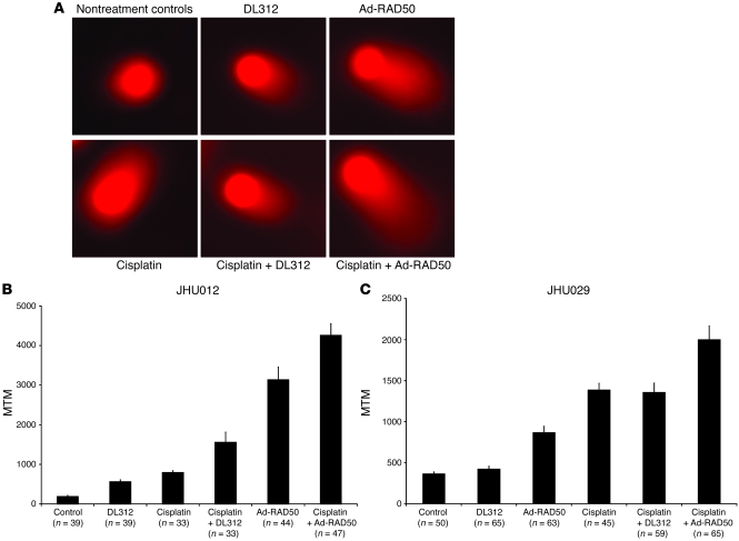
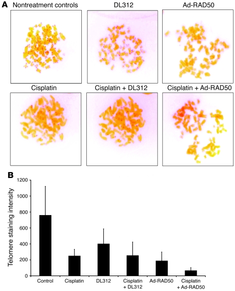
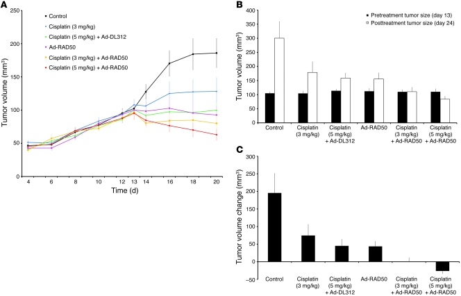
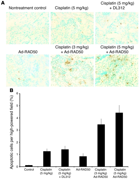
Platinum-based drugs that induce DNA damage are commonly used first-line chemotherapy agents for testicular, bladder, head and neck, lung, esophageal, stomach, and ovarian cancers. The inherent resistance of tumors to DNA damage often limits the therapeutic efficacy of these agents, such as cisplatin. An enhanced DNA repair and telomere maintenance response by the Mre11/Rad50/Nbs1 (MRN) complex is critical in driving this chemoresistance. We hypothesized therefore that the targeted impairment of native cellular MRN function could sensitize tumor cells to cisplatin. To test this, we designed what we believe to be a novel dominant-negative adenoviral vector containing a mutant RAD50 gene that significantly downregulated MRN expression and markedly disrupted MRN function in human squamous cell carcinoma cells. A combination of cisplatin and mutant RAD50 therapy produced significant tumor cytotoxicity in vitro, with a corresponding increase in DNA damage and telomere shortening. In cisplatin-resistant human squamous cell cancer xenografts in nude mice, this combination therapy caused dramatic tumor regression with increased apoptosis. Our findings suggest the use of targeted RAD50 disruption as what we believe to be a novel chemosensitizing approach for cancer therapy in the context of chemoresistance. This strategy is potentially applicable to several types of malignant tumors that demonstrate chemoresistance and may positively impact the treatment of these patients.


Figures

Figure 1
Figure 1. Native MRN expression in head and neck SCC cells.
Native expression of MRE11, RAD50, and NBS1 was confirmed in both JHU012 and JHU029 SCC cells by Western blot. Lane 1, JHU029 cells; lane 2, JHU012 cells; lane 3, MagicMark Western Protein Standard with markers of 20, 30, 40, 50, 60, 80, 100, 120, and 220 kDa from bottom to top. Lanes were run on the same gel but were noncontiguous as indicated by the vertical white lines.
Figure 2
Figure 2. Construction and function of the Ad-RAD50 vector.
(A) A 326–base pair mutant RAD50 hook fragment from the wild-type RAD50 gene was cloned into a recombinant adenovirus vector to construct Ad-RAD50. The amino acid sequence of the integrated construct (residues 631–739) is highlighted in black, with the zinc hook region marked in yellow. PA, polyadenylation site. (B) Both the wild-type RAD50 protein and the mutant RAD50 protein were detected by Western blot in JHU012 tumor cells that were infected with Ad-RAD50 adenovirus. Lane 1, MagicMark Western Protein Standard; lane 2, JHU012 cells with no treatment; lane 3, JHU012 cells infected with Ad-RAD50.
Figure 3
Figure 3. Mutant RAD50 protein interacts with endogenous RAD50 and downregulates the MRN complex.
(A) Anti-MRE11 antibody coimmunoprecipitated wild-type RAD50 in all cells, suggesting a direct interaction between these proteins. Mutant RAD50 was only coimmunoprecipitated in infected cells treated with zinc, indicating that the interaction between mutant RAD50 and the wild-type MRN complex is mediated by zinc-dependent dimerization of RAD50 hook domains and not direct interactions between mutant RAD50 and MRE11. All lanes shown were run simultaneously on a single gel as contiguous lanes. (B) Multiple MRN nuclear foci are seen at sites of DNA damage in JHU012 cells treated with Ad-RAD50 and cisplatin. Original magnification, ×63. There is spatial overlap of the signals corresponding to wild-type RAD50 (blue) and wild-type MRE11 (red), highly suggestive of a binding interaction between these proteins. (C) Nuclear foci from JHU012 cells treated with Ad-RAD50 and cisplatin (original magnification, ×63) demonstrate spatial overlap of the wild-type (red) and mutant RAD50 (blue) proteins, further suggesting direct binding between these proteins. (D) Downregulation of wild-type MRE11 (P < 0.005), RAD50 (P < 0.001), and NBS1 proteins (P < 0.04) is seen after infection of JHU012 cells with Ad-RAD50 relative to noninfected controls. (Mean optical density of Western blot bands ± SEM is shown.) This is attributable to the demonstrated dimerization between mutant and wild-type RAD50 and inability of the mutant protein to directly bind MRE11. Consequently, only a single MRE112/RAD502 heterotetramer can form around the wild-type RAD50 with no complimentary heterotetramer assembling around mutant RAD50.
Figure 4
Figure 4. Ad-RAD50 enhances cisplatin-induced suppression of SCC cell growth.
(A) Combining Ad-RAD50 and cisplatin in JHU012 SCC cells produced a reduction in cell growth within 48 hours and a persistent suppression of cell proliferation relative to all other groups (P < 0.01). DL312 indicates DL312 control virus. (B) The addition of Ad-RAD50 to cisplatin treatment in JHU029 cells produced the greatest reduction in cell growth versus all other groups (P < 0.01). Cells were cloned from parental cell lines. Mean optical density ± SEM is shown.
Figure 5
Figure 5. Ad-RAD50 enhances cisplatin-induced DNA damage.
(A) Neutral comet assay was used to quantify the level of DNA DSB damage in JHU012 (shown) SCC cells. Damaged, fragmented DNA migrates toward the anode, producing a comet tail. Original magnification, ×20. (B) MTM as measured in the JHU012 cell line. The MTM is a correlate of DNA DSB damage. MTM ± SEM is shown. Ad-RAD50 combined with cisplatin increased the number of DNA DSBs by 437% relative to cisplatin monotherapy (P < 0.001) and by 173% compared with Ad-RAD50 alone (P < 0.02), suggesting that the enhanced cytotoxic effect of combined therapy is secondary to increased DNA DSB damage. (C) MTM as measured in the JHU029 cell line. MTM ± SEM is shown. Ad-RAD50 combined with cisplatin significantly increased DSB damage compared with all other groups (P < 0.003).
Figure 6
Figure 6. Ad-RAD50 and cisplatin combination treatment enhances telomere shortening in JHU012 SCC cells.
(A) PNA FISH stains telomeres with a red signal, the intensity of which is proportional to telomere length. Original magnification, ×100. (B) Software-based quantification of telomere staining intensity indicated that combining Ad-RAD50 with cisplatin markedly attenuated telomere length by 73.9% versus cisplatin alone and by 65.2% versus Ad-RAD50 (P < 0.01). Telomere shortening beyond a critical threshold induces apoptosis, contributing to the cytotoxic effect of combination therapy in tumor cells. Mean ± SEM is shown.
Figure 7
Figure 7. Ad-RAD50 chemosensitizes JHU012 human head and neck cancer to cisplatin in vivo.
(A) Externally measured tumor volume ± SEM from the time of initial tumor palpability on day 4, to treatment initiation on day 13, and through the treatment period to day 20. (B) Mean internally measured tumor volume ± SEM immediately prior to treatment on day 13 and at the time of animal sacrifice on day 24. (C) Mean internally measured change in tumor volume ± SEM. During the 11 day period between treatment initiation and animal sacrifice, a single administration of Ad-RAD50 combined with a single intraperitoneal dose of cisplatin, at either 3 mg/kg or 5 mg/kg, was able to significantly enhance the therapeutic efficacy of cisplatin (3 mg/kg or 5 mg/kg) monotherapy. For Ad-RAD50 with cisplatin (3 mg/kg), P < 0.001 vs. control; P < 0.05 vs. cisplatin (3 mg/kg) and cisplatin (5 mg/kg) with DL312; and P < 0.05 vs. Ad-RAD50 alone. For Ad-RAD50 with cisplatin (5 mg/kg), P < 0.001 vs. control, cisplatin (3 mg/kg), cisplatin (5 mg/kg) with DL312, and Ad-RAD50 alone.
Figure 8
Figure 8. Quantification of apoptosis in human head and neck cancer tumors.
(A) TUNEL staining was performed on tumor SCC samples after tumor harvest. A selection of 1 high-powered field (original magnification, ×40) from each treatment group is shown. (B) Mean percentage apoptosis per high-powered field ± SEM is shown. The combination of Ad-RAD50 and cisplatin enhanced induction of apoptosis relative to all other groups (P < 0.001). This is consistent with the tumor volume reduction seen in the combination therapy groups.

Similar articles

Cited by

References

    1. Kelland L. The resurgence of platinum-based cancer chemotherapy. Nat. Rev. Cancer. 2007;7:573–584. doi: 10.1038/nrc2167. - DOI - PubMed
    1. Chu G. Cellular responses to cisplatin. The roles of DNA-binding proteins and DNA repair. J. Biol. Chem. 1994;269:787–790. - PubMed
    1. Perez R.P. Cellular and molecular determinants of cisplatin resistance. Eur. J. Cancer. 1998;34:1535–1542. doi: 10.1016/S0959-8049(98)00227-5. - DOI - PubMed
    1. Rodenburg C.J., Cleton F.J. Chemotherapy in advanced ovarian cancer. J. Cancer Res. Clin. Oncol. 1984;107:99–105. doi: 10.1007/BF00399380. - DOI - PubMed
    1. Jacobs C., et al. A phase III randomized study comparing cisplatin and fluorouracil as single agents and in combination for advanced squamous cell carcinoma of the head and neck. J. Clin. Oncol. 1992;10:257–263. - PubMed

Publication types

MeSH terms