

Biomarkers for epithelial-mesenchymal transitions
- PMID: 19487819
- PMCID: PMC2689132
- DOI: 10.1172/JCI36183
Biomarkers for epithelial-mesenchymal transitions
Abstract
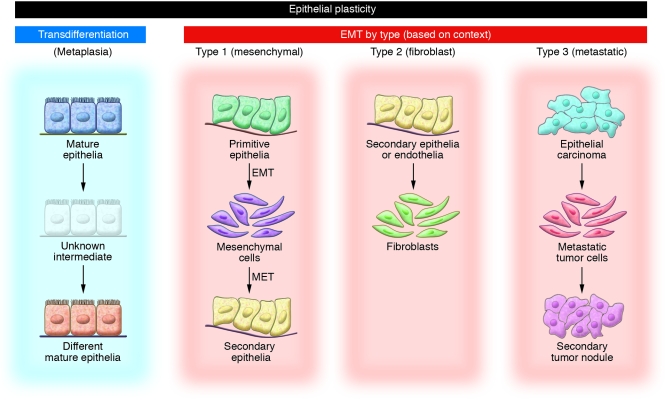
Somatic cells that change from one mature phenotype to another exhibit the property of plasticity. It is increasingly clear that epithelial and endothelial cells enjoy some of this plasticity, which is easily demonstrated by studying the process of epithelial-mesenchymal transition (EMT). Published reports from the literature typically rely on ad hoc criteria for determining EMT events; consequently, there is some uncertainty as to whether the same process occurs under different experimental conditions. As we discuss in this Personal Perspective, we believe that context and various changes in plasticity biomarkers can help identify at least three types of EMT and that using a collection of criteria for EMT increases the likelihood that everyone is studying the same phenomenon - namely, the transition of epithelial and endothelial cells to a motile phenotype.
Figures
References
-
- Mazzarello P. A unifying concept: the history of cell theory. Nat. Cell Biol. 1999;1:E13–E15. - PubMed
-
- Blau H.M., Blakely B.T. Plasticity of cell fate: insights from heterokaryons. Semin. Cell Dev. Biol. 1999;10:267–272. - PubMed
-
- Rizzino A. A challenge for regenerative medicine: proper genetic programming, not cellular mimicry. Dev. Dyn. 2007;236:3199–3207. - PubMed
-
- Neilson E.G. Plasticity, nuclear diapause, and a requiem for the terminal differentiation of epithelia. J. Am. Soc. Nephrol. 2007;18:1995–1998. - PubMed
Publication types
MeSH terms
Substances
Grants and funding
LinkOut - more resources
Full Text Sources
Other Literature Sources
