

Review
doi: 10.1172/JCI38019.
Epub 2009 Jun 1.
Epithelial-mesenchymal transitions: the importance of changing cell state in development and disease
Affiliations
- PMID: 19487820
- PMCID: PMC2689100
- DOI: 10.1172/JCI38019
Item in Clipboard
Review
Epithelial-mesenchymal transitions: the importance of changing cell state in development and disease
J Clin Invest.
2009 Jun.
Abstract
The events that convert adherent epithelial cells into individual migratory cells that can invade the extracellular matrix are known collectively as epithelial-mesenchymal transition (EMT). Throughout evolution, the capacity of cells to switch between these two cellular states has been fundamental in the generation of complex body patterns. Here, we review the EMT events that build the embryo and further discuss two prototypical processes governed by EMT in amniotes: gastrulation and neural crest formation. Cells undergo EMT to migrate and colonize distant territories. Not surprisingly, this is also the mechanism used by cancer cells to disperse throughout the body.
Figures
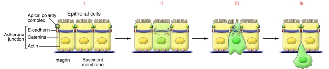
(i) Normal epithelial cells contain adherens junctions composed of E-cadherin together with catenins and actin rings. Tight junctions are associated with apical polarity complexes, while integrins interact with components of the basal membrane. (ii) Loss of cell-cell adhesion. EMT inducers repress the transcription of the genes encoding the components of both adherens and tight junctions, inducing the loss of cell polarity. E-cadherin is internalized and targeted for degradation. (iii) Breakdown of the basal membrane and apical constriction. Profound cytoskeletal remodeling will favor cell delamination by inducing apical constriction and disorganization of the basal membrane. (iv) Cell delamination and invasion. Expression of integrin receptors and continued activation of metalloproteases favors migration through the extracellular matrix and invasion of adjacent tissues.
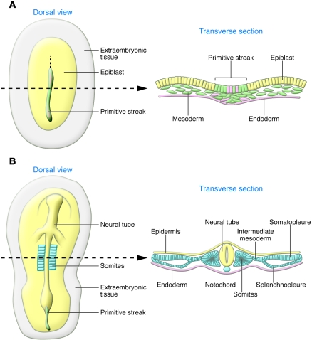
Schematic representation of chick embryos as representative of amniotes (birds, reptiles, and mammals). Dorsal views and transverse sections taken at the level of the dotted lines. (A) The embryonic layers are defined during gastrulation. Mesodermal (green) and endodermal cells (pink) are internalized at the primitive streak through a process of EMT, while ectodermal cells remain epithelial (yellow). (B) The mesodermal cells condense to form various derivatives (blue) along the medio-lateral axis of the embryo. The axial mesoderm gives rise to the notochord; paraxial mesoderm epithelializes through a process of MET to form the somites; the intermediate mesoderm will later form the urogenital system; and the lateral mesoderm condenses to form somatopleure and splanchnopleure. The ectodermal cells contribute to the neural tube or the epidermis. The endoderm is shown in pink.
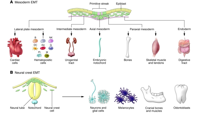
(A) Epiblast cells that internalize at gastrulation give rise to different mesodermal and endodermal populations from which a variety of cell types form. Embryonic cells undergoing EMT are shown in green. Pt, platelets; B, T, and NK, lymphocytes; G, granulocytes; M, macrophages. (B) In turn, the neural crest delaminates from the dorsal neural tube and will generate neurons of the peripheral nervous system, glial and satellite cells, pigment cells, odontoblasts, and the craniofacial cartilage, as well as other cell types.
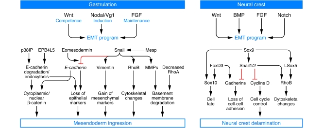
Signaling molecules of the TFG-β superfamily (Nodal, Vg1, and BMPs), together with Wnt and FGF, initiate the formation of the primitive streak and operate at the neural folds to drive the ingression of the mesendoderm and the delamination of the neural crest, respectively. Some downstream targets are also conserved, such as the Snail genes. While Snail factors are key regulators of the EMT program during gastrulation, the coordinated induction of several transcription factors is required to control the robust program of neural crest delamination. EPB4L5, FERM and actin-binding domain–containing band 4.1 superfamily member; p38IP, p38-interacting protein; Rho, members of the Rho family of small GTPases.
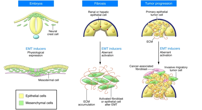
EMTs (cells in green) occur during normal embryonic development, such as during neural crest cell delamination from the dorsal neural tube and mesendoderm ingression from the primitive streak. While EMT inducers are usually maintained in a silent state in the adult, they are reactivated during organ fibrosis and at the invasive front of human carcinomas during tumor progression.
References
-
- Thiery J.P., Sleeman J.P. Complex networks orchestrate epithelial-mesenchymal transitions. Nat. Rev. Mol. Cell Biol. 2006;7:131–142. - PubMed
-
- Hay, E.D. 1968. Organization and fine structure of epithelium and mesenchyme in the developing chick embryo. In Epithelial-mesenchymal interactions. R. Fleischmajer and R.E. Billingham, editors. Williams & Wilkins. Baltimore, Maryland, USA. 31–55.
-
- Barrallo-Gimeno A., Nieto M.A. The Snail genes as inducers of cell movement and survival: implications in development and cancer. Development. 2005;132:3151–3161. - PubMed
-
- Peinado H., Olmeda D., Cano A. Snail, Zeb and bHLH factors in tumor progression: an alliance against the epithelial phenotype? Nat. Rev. Cancer. 2007;7:415–428. - PubMed
-
- Moreno-Bueno G., Portillo F., Cano A. Transcriptional regulation of cell polarity in EMT and cancer. Oncogene. 2008;27:6958–6969. - PubMed
