. 2009 Jan-Feb;75(1):43-9.
doi: 10.1016/s1808-8694(15)30830-2.

Understanding the anatomy of ears from guinea pigs and rats and its use in basic otologic research

Affiliations

Understanding the anatomy of ears from guinea pigs and rats and its use in basic otologic research

Agnes Afrodite Sumarelli Albuquerque et al. Braz J Otorhinolaryngol. 2009 Jan-Feb.

Abstract

The use of animal samples is important in otologic research and understanding the anatomy of their ears help make proper use of them in research projects.

Aim: to study guinea pig's and rat's ears under light microscopy(LM) and scanning electron microscopy(SEM) and understand their anatomical advantages in basic otologic research.

Materials and methods: The temporal bones, tympanic bullas and cochleas from three albino guinea pigs and rats were photographed and analyzed under LM and SEM.

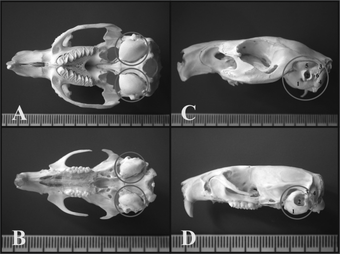
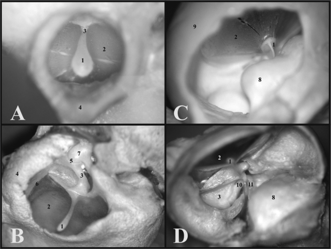
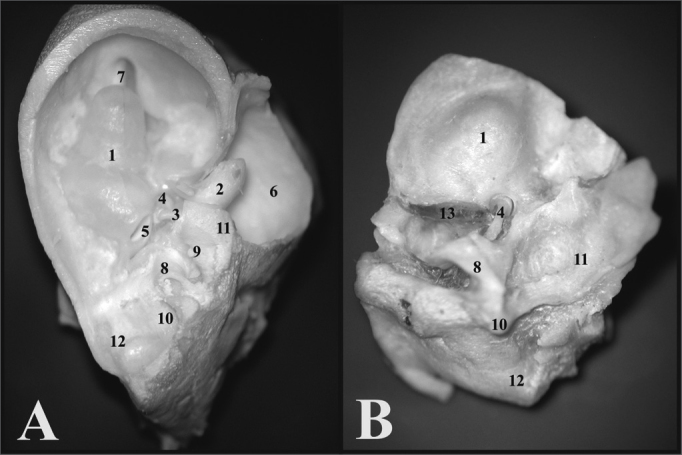
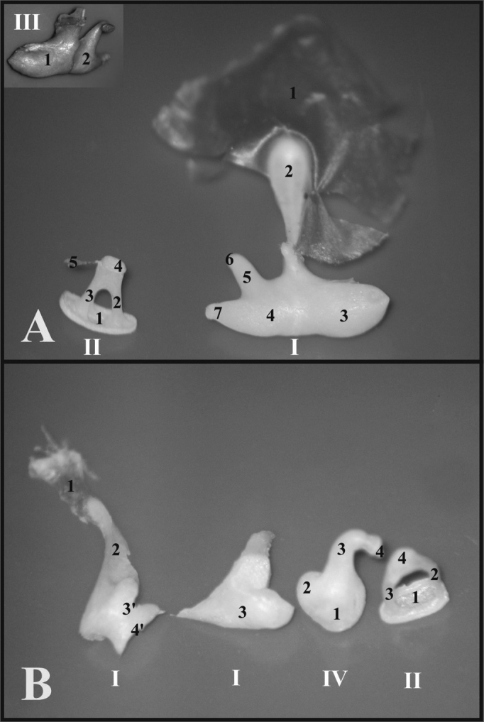
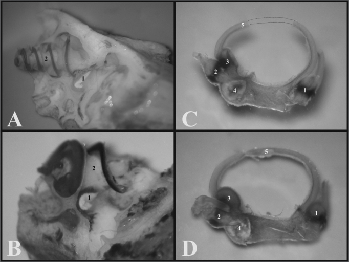
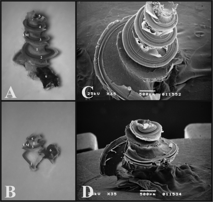
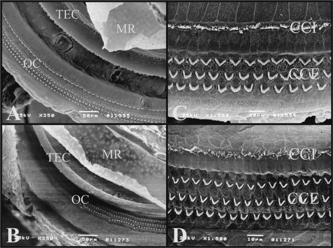
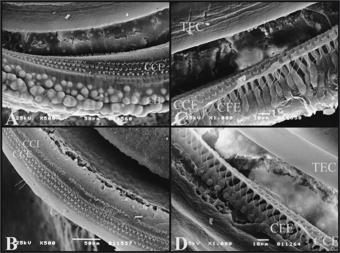
Results: Rats aren't as simple to handle as guinea pigs, and often present with otitis media. Rats have a fragile junction of the tympanic bulla, two and half turns in the cochlea, and their tympanic membranes do not seal off the entire external auditory canal. Guinea pigs have full bullas, their incus and malleus are fused and they have three and half cochlear turns. Under SEM, guinea pigs and rats have Tectori Membrane, Raissner's Membrane and the Organ of Corti. Only guinea pigs have Hensen's Cells.

Conclusion: Guinea pigs were considered easy to handle for microdissection purposes because of the size and robustness of their temporal bones, and for surgical experiments involving the stapes, the oval window and the tympanic membrane. Under SEM there are similarities guinea pigs and rats, and both can be used in inner ear studies.


Figures

Figure 1
Figure 1
A) Guinea pig’s skull base showing the tympanic bullas (lower view) (millimeter scale); B) Rat’s skull base showing the tympanic bullas (lower view) (millimeter scale); C) Lateral left-side view of a young guinea pig’s skull (millimeter scale); D) Lateral left-side view of a young-adult rat’s skull - (millimeter scale). 1 - Bulla; 2 - External auditory canal; 3 - Squamous bone; 4 - Posterior air Sinus notch; 5 - Anterior air sinus; 6 - Para-occipital apophysis.
Figure 2
Figure 2
A) View through the external auditory canal of a young guinea pig (microphotography, 60X magnification); B) View through the external auditory canal of a young-adult rat (microphotography, 75X magnification); C) Inside view of the auditory cleft in a young guinea pig (inside the left tympanic bulla) (microphotography, 58X magnification); D) Inside view of the auditory cleft of a young-adult rat (inside the left tympanic bulla) (microphotography, 75X magnification). 1 - Malleus handle; 2 - Tympanic membrane; 3 - Malleus (neck); 4 - External auditory canal; 5 - Incus-malleus joint; 6 - Tympanic ring; 7 - Incus; 8 - Cochlea with bony cover; 9 - Bulla; 10 - Long process of the incus; 11 - Stapes.
Figure 3
Figure 3
A) View inside the tympanic bulla of a young guinea pig with ossicles and cochlea with the bony cover (microphotography, 15X magnification); B) View inside the tympanic bulla of the young-adult rat with the stapes and the cochlea with the bony cover (microphotography, 28X magnification). 1 - Cochlea; 2 - Malleus; 3 - Long process of the incus; 4 - Stapes footplate over the oval window (stapes); 5 - Round window; 6 - Mastoid; 7 - Eustachian tube opening; 8 - Lateral semicircular canal projection; 9 - Facial nerve canal bony projection; 10 - Posterior semicircular canal projection; 11 - Superior semicircular canal projection; 12 - Posterior air sinus projection; 13 - Carotid artery.
Figure 4
Figure 4
A) Set of ossicles of a young guinea pig. Detailed view of the malleus-incus joint (III) (microphotography, 58X magnification); B) Set of ossicles from a young-adult rat (microphotography, 60X magnification). I - Malleus - Incus 1 - Tympanic membrane; 2 - Malleus handle; 3 - Head of the malleus; 3’ - Neck of the malleus; 4 - Incus (fused to the malleus); 4’ - Joint surface of the malleus-incus; 5 - Long process of the incus; 6 - Lenticular process of the incus; 7 - short process of the incus II - Stapes 1 - Footplate of the incus; 2 - anterior crus of the stapes; 3 - Stapes posterior crus; 4 - Stapes Capitulum; 5 - Stapes muscle tendon III - Malleus-incus joint 1 - Malleus; 2 - Incus IV - Incus 1 - Incus-Malleus joint surface; 2 - Short process of the incus; 3 - Long process of the incus; 4 - Lenticular process of the incus.
Figure 5
Figure 5
A) Internal wall of the left labyrinth from a young guinea pig showing the vestibule (microphotography, 55X magnification); B) Internal wall of the left labyrinth of a young-adult rat, showing the vestibule (microphotography, 60X magnification); C) Left membranous labyrinth of a young guinea pig (microphotography, 48X magnification); D) left membranous labyrinth from a young-adult rat (microphotography, 48X magnification). A and B) 1 - Saccule 2 - Cochlea C and D) 1 - posterior semicircular canal ampulla; 2 - superior semicircular canal ampulla; 3 - Lateral semicircular canal ampulla; 4 - Utricle; 5 - Lateral semicircular canal
Figure 6
Figure 6
A) Cochlea from a young guinea pig, showing the cochlear turns with the membranous labyrinth (ML) (Microphotography, 44X magnification); B) Cochlea from a young-adult rat, showing the cochlear turns with the membranous labyrinth (ML) (Microphotography, 44X magnification); C) Guinea pig cochlea seen under scanning electron microscope (scanning electron micrograph, 35X magnification); D) Rat cochlea seen under scanning electron microscopy (scanning electromicrography, 35X magnification). 1 - Modiolus; 2 - Basal turn (E1); 3 - Turn 2 (E2); 4 - Turn 3 (E3); 5 - Apical turn; 6 - Cochlear nerve.
Figure 7
Figure 7
A) Guinea pig cochlea turn showing the Raissner’s Membrane (RM), Tectorial Membrane (TEC), Organ of Corti (OC) (Scanning electromicrography, 350X Magnification (TEC), Organ of Corti (OC) (Scanning electromicrography of the cochlear basal turn with the outer hair cells (CCE) and inner hair cells (CCI) (scanning electron microscopy, 1500X magnification); D) Organ of Corti from the basal turn of the rat’s cochlea with the Outer Hair Cells (CCE) and Inner Hair Cells (CCI) (scanning electron microscopy, 1500X magnification).
Figure 8
Figure 8
A) 3rd cochlear turn of the guinea pig showing the Hansen Cells (CH) (scanning electron micrograph, 500X magnification); B) 2nd cochlear turn from a rat, showing the lack of Hansen Cells definition (Scanning electron microscopy, 500X magnification); C) Guinea pig’s cochlea basal turn showing the external phalanx cells (CFE) (Deiters) (scanning electron micrography, 1000X magnification); D) Rat’s cochlea basal turn showing the external phalanx cells (CFE) (Deiters) (Scanning electron micrography, 1000X magnification).

References

    1. Schanaider A, Silva PC. uso de animais em cirurgia experimental. Acta Cir Bras. 2004;19(4):441–447.
    1. Oliveira JAA. Audio-Vestibular toxicity of drugs. CRC Press; Florida: 1989. p. 560.
    1. Santos PF, et al. Achados otomicrocópicos e histológicos da miringoes-clerose induzida em ratos: estudo crítico de um modelo experimental. Rev Bras Otorrinolaringol. 2005;71(5):668–674.
    1. Judkins RF, LI H. Surgical anatomy of the rat middle ear. Otolaryngol Head Neck Surg. 1997;117(5):438–447. - PubMed
    1. Pinilla M, Ramírez-Camacho R, Jorge E, Trinidad A, Vergara J. Ventral approach to the rat middle ear for otologic research. Otolaryngol Head Neck Surg. 2001;124(5):515–517. - PubMed

MeSH terms

LinkOut - more resources