. 2009:544:637-53.
doi: 10.1007/978-1-59745-483-4_41.

Determining DNA sequence specificity of natural and artificial transcription factors by cognate site identifier analysis

Affiliations

Determining DNA sequence specificity of natural and artificial transcription factors by cognate site identifier analysis

Mary S Ozers et al. Methods Mol Biol. 2009.

Abstract

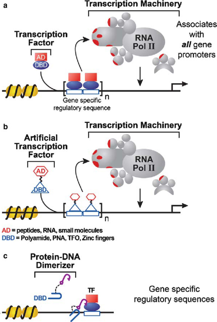
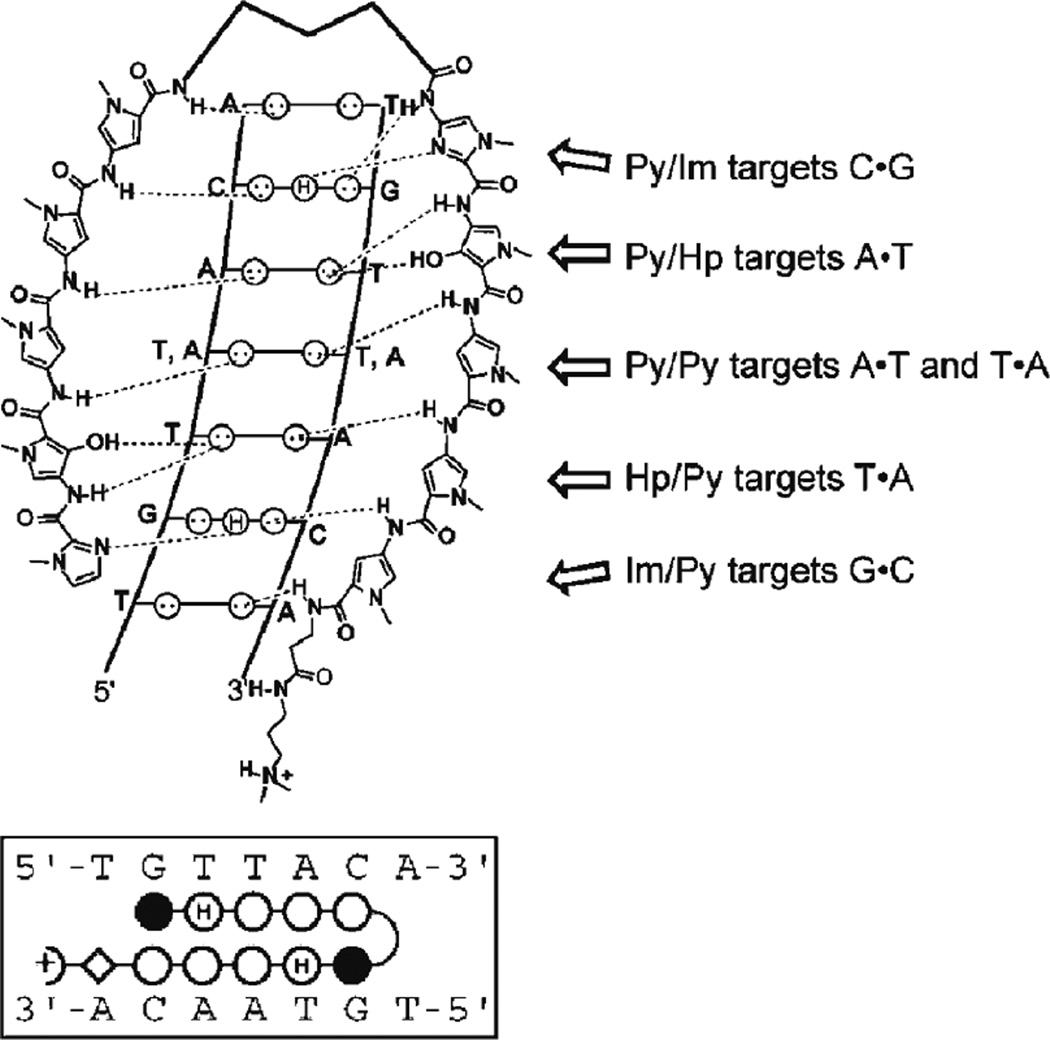
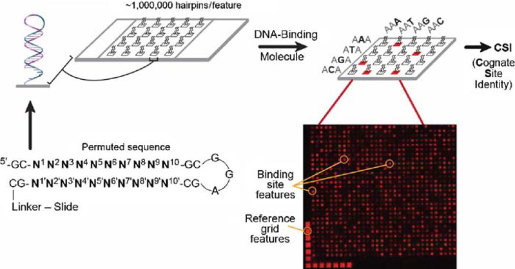
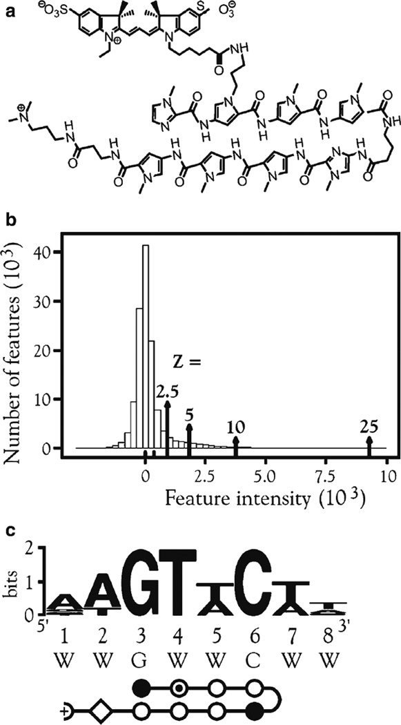
Artificial transcription factors (ATFs) are designed to mimic natural transcription factors in the control of gene expression and are comprised of domains for DNA binding and gene regulation. ATF domains are modular, interchangeable, and can be composed of protein-based or nonpeptidic moieties, yielding DNA-interacting regulatory molecules that can either activate or inhibit transcription. Sequence-specific targeting is a key determinant in ATF activity, and DNA-binding domains such as natural zinc fingers and synthetic polyamides have emerged as useful DNA targeting molecules. Defining the comprehensive DNA binding specificity of these targeting molecules for accurate manipulations of the genome can be achieved using cognate site identifier DNA microarrays to explore the entire sequence space of binding sites. Design of ATFs that regulate gene expression with temporal control will generate important molecular tools to probe cell- and tissue-specific gene regulation and to function as potential therapeutic agents.


Figures

Fig. 1
Fig. 1
Role of ATFs and protein–DNA dimerizers in transcription. (a) Transcription factors are modular proteins composed of a DNA-binding domain (DBD) that recognizes gene-specific regulatory sequences and an activation domain (AD) that recruits RNA polymerase II and/or associated proteins. (b) Artificial transcription factors (ATFs) can be created using transcriptional activation domains such as VP16 or small molecules and DBDs such as protein-based DNA-binding modules or synthetic molecules including polyamides, triplex-forming oligonucleotides (TFOs), or peptide nucleic acids (PNAs). (c) Protein–DNA dimerizers contain a DBD linked to a molecular “hook” such as a short peptide or small molecule, which facilitates binding of a natural transcription factor at an adjacent DNA site.
Fig. 2
Fig. 2
Polyamide pairing rules. (a) Polyamides target DNA based on a set of pairing rules. An Im/Py ring pair recognizes G·C; Py/Py pair targets A·T or T·A; and Py/Hp binds to A·T (18). (b) Polyamides can be represented by the following abbreviations and symbols:Im, N-methylimidazole, filled circle; Py, N-methylpyrrole, open circle; Hp, hydroxypyrrole, open circle with H; β, β-alanine, diamond; Dp, dimethylaminopripylamide, half circle with positive charge; γor turn, γ-aminobutyric acid.
Fig. 3
Fig. 3
Illustration of a CSI microarray. Every permutation of a 10 bp (N1–N10) sequence is displayed in a hairpin probe containing a GC stem and GGA turn. The array is incubated with a fluorescently labeled DNA-binding molecule. The fluorescent features are identified and used to determine DNA-binding preferences.
Fig. 4
Fig. 4
Binding of PA1. (a) Structure of Cy3-conjugated polyamide PA1 (ImPy*PyPy-γ-ImPyPyPy-β-Dp). (b) Histogram of averaged intensities of all replicate features. Z scores (see Methods) are noted. (c) Logo obtained from top Z-score bin of 25. Abbreviations: Py*, N-methylpyrrole ring with Cy3 dye attached, open circle with inner dot; W = A or T.
Fig. 5
Fig. 5
Polyamide–Exd cooperative complex. (a) Schematic of polyamide–Exd cooperative complex on DNA. (b) Logo (–28) obtained from CSI analysis of polyamide–Exd binding. Boxed sequence displays binding sites for Exd and polyamide. (c) The polyamide-peptide conjugate at 50 nM was incubated with increasing concentrations of Exd (nM). Arrows indicate free DNA (lower arrow) and DNA–polyamide complex (upper arrow). Boxed sequence denotes the Exd and polyamide-binding sites. (d) Molecular modeling (29, 30) of Exd (green) and polyamide (blue) bound to consensus DNA site, with peptide hook (brown) and linker (red). Models were obtained by aligning crystal structures of the DNA complexed with Exd or hairpin polyamide (Protein Data Bank files 1B8I and 1M18).

References

    1. Ren B, Robert F, Wyrick JJ, Aparicio O, Jennings EG, Simon I, Zeitlinger J, Schreiber J, Hannett N, Kanin E, Volkert TL, Wilson CJ, Bell SP, Young RA. Genome-wide location and function of DNA binding proteins. Science. 2000;290:2306–2309. - PubMed
    1. Venter JC, et al. The sequence of the human genome. Science. 2001;291:1304–1351. - PubMed
    1. Wells J, Graveel CR, Bartley SM, Madore SJ, Farnham PJ. The identification of E2F1-specific target genes. Proc. Natl Acad. Sci. U. S. A. 2002;99:3890–3895. - PMC - PubMed
    1. Horak CE, Mahajan MC, Luscombe NM, Gerstein M, Weissman SM, Snyder M. GATA-1 binding sites mapped in the beta-globin locus by using mammalian ChIP-chip analysis. Proc. Natl Acad. Sci. U. S. A. 2002;99:2924–2929. - PMC - PubMed
    1. Martone R, Euskirchen G, Bertone P, Hartman S, Royce TE, Luscombe NM, Rinn JL, Nelson FK, Miller P, Gerstein M, Weissman S, Snyder M. Distribution of NF-kappaB-binding sites across human chromosome 22. Proc. Natl Acad. Sci. U. S. A. 2003;100:12247–12252. - PMC - PubMed

Publication types

LinkOut - more resources