. 2009;53(4):535-40.
doi: 10.1387/ijdb.092855bp.

Developmental expression and regulation of the chemokine CXCL14 in Xenopus

Affiliations

Developmental expression and regulation of the chemokine CXCL14 in Xenopus

Byung-Yong Park et al. Int J Dev Biol. 2009.

Abstract

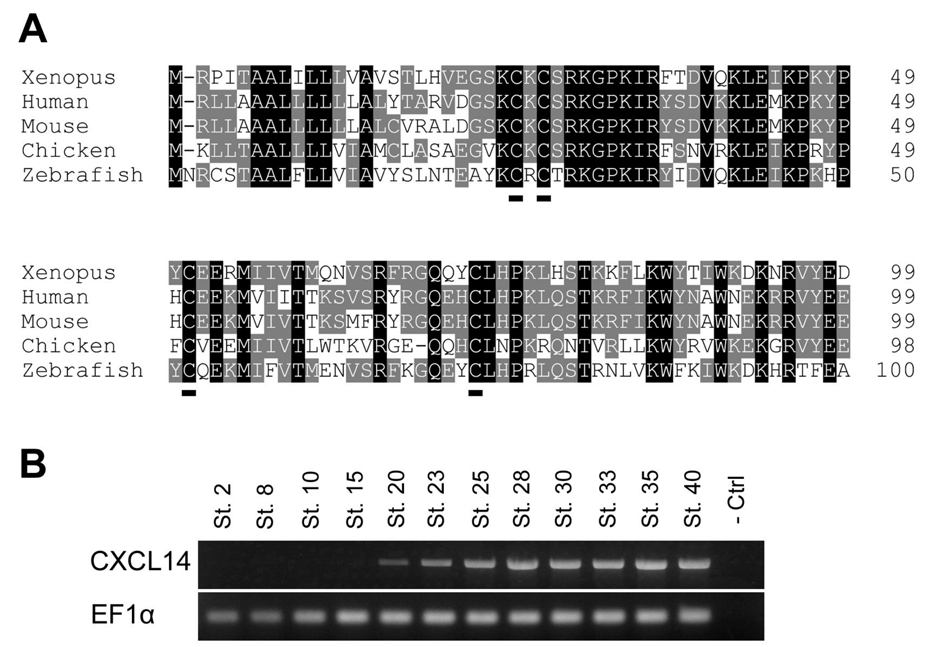
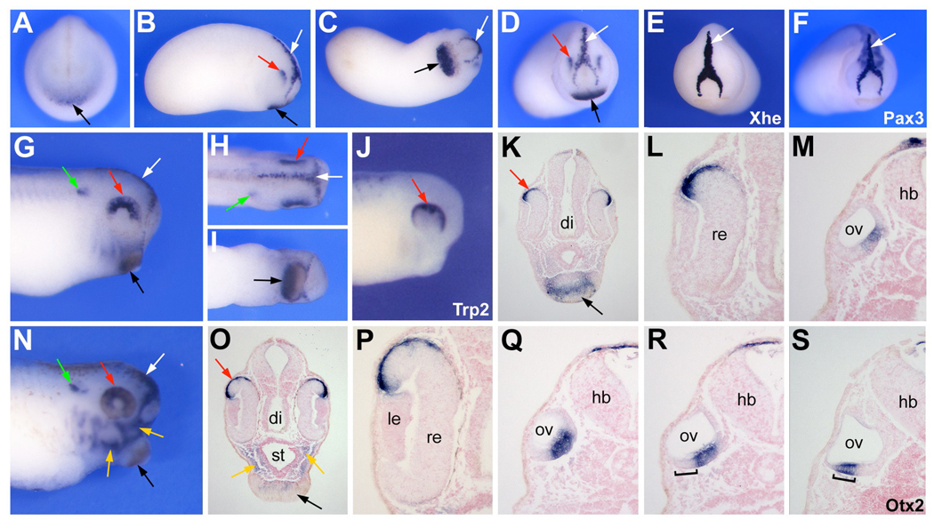
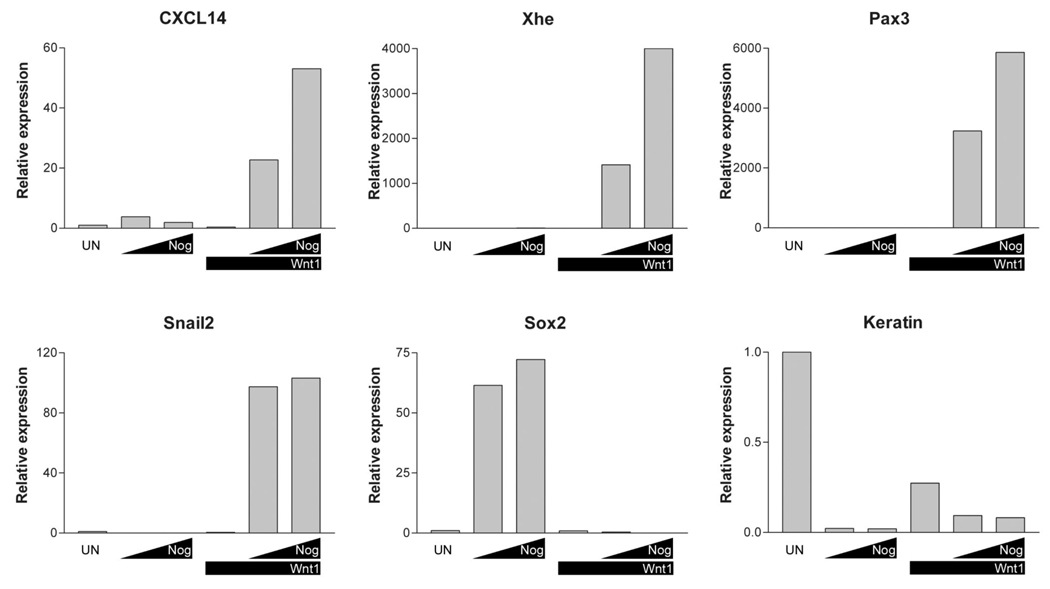
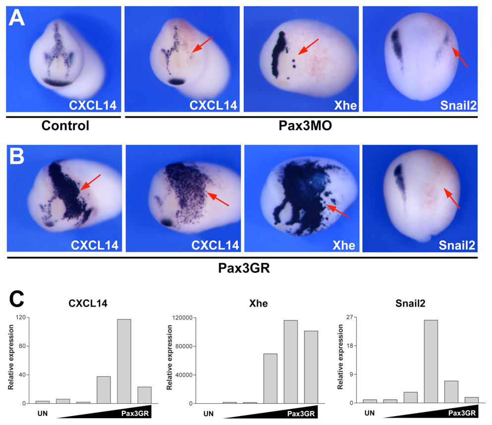
Chemokines are a family of proteins originally identified for their activity promoting the recruitment of leukocytes to inflammatory sites. Recent evidence indicates that chemokines and their receptors may also regulate key developmental processes. In this paper we report the expression and regulation of the chemokine CXCL14 during Xenopus laevis embryogenesis. CXCL14 is first detected in several ectoderm derivatives, the dorsal aspect of the retina, the cement gland and the hatching gland. Later in development, additional domains of expression include the head mesenchyme and the medial ventral aspect of the otic vesicle. CXCL14 expression in the ectoderm is regulated by both Bmp and canonical Wnt signaling. In the hatching gland CXCL14 is co-expressed with the transcription factor Pax3. Using gain of function and knockdown approaches in whole embryos and animal explants we show that Pax3 is both necessary and sufficient for CXCL14 expression in this domain of the ectoderm.


Figures

Figure 1
Figure 1. Sequence and developmental expression of Xenopus CXCL14
(A) Deduced amino acid sequences from human, mouse, chicken and zebrafish CXCL14 were aligned using GeneDoc. Conserved amino acids in all five species or in at least two species are highlighted in black and grey, respectively. The conserved cysteine residues, signature motif of this class of molecules, are underlined. (B) RT-PCR analysis of the developmental expression of CXCL14. Stages are according to Nieuwkoop and Faber (1967). EF1α is shown as a loading control.
Figure 2
Figure 2. Expression of CXCL14 by whole-mount in situ hybridization
(A) Onset of CXCL14 expression at stage 20 in the prospective cement gland anterior to the neural plate (black arrow). Frontal view, dorsal to top. (B–D) At stage 23 CXCL14 is detected in part of the cement gland (black arrows), the dorsal anterior portion of the optic vesicle (red arrows) and the hatching gland (white arrows). (B) Lateral view, dorsal to top, anterior to right. (C) Ventral view, anterior to right. (D) Frontal view, dorsal to top. The expression of Xhe (E) and Pax3 (F) in the hatching gland (white arrows) at this stage are shown for comparison. (E–F) Frontal view, dorsal to top. (G–I) CXCL14 expression at stage 28. In addition to the cement gland (black arrows), optic vesicle (red arrows) and hatching gland (white arrows), CXCL14 is also detected in the otic vesicle (green arrows). (G) Lateral view, dorsal to top, anterior to right. (H) Dorsal view, anterior to right. (H) Ventral view, anterior to right. (J) The expression pattern of Trp2 in the pigmented epithelium of the retina is similar to CXCL14 expression in the developing eye (red arrow). Lateral view, dorsal to top, anterior to right. (K–L) Transverse section highlights the expression of CXCL14 expression in the pigmented epithelium of the retina (red arrow). Panel (L) is a higher magnification view of panel (K). CXCL14 expression is restricted to the ventral medial aspect of the otic vesicle (M). (N) At stage 35, CXCL14 expression persists in the same domains and is also detected in the head mesenchyme (yellow arrows). Lateral view, dorsal to top, anterior to right. (O–R) Transverse sections showing the expression of CXCL14 in the head mesenchyme and the cement gland (O), pigmented epithelium of the retina (O–P) and otic vesicle (Q–R). Panel (Q) corresponds to a section posterior to section shown in panel (R). For comparison at this stage Otx2 (bracket) is expressed in the ventral aspect of the otic vesicle (S) a domain that does not overlap with CXCL14 (R). di, diencephalon; hb, hindbrain; le, lens; ov, otic vesicle; re, retina; st, stomodeum.
Figure 3
Figure 3. CXCL14 expression in the ectoderm is regulated by both Bmp and canonical Wnt signaling
Embryos at the 2-cell stage were injected in the animal pole with two distinct doses of Noggin (N) mRNA (0.4 ng and 1.0 ng) alone or in combination with Wnt1 (+W) mRNA (0.1 ng). Animal explants were dissected at the blastula stage, cultured for 10 hours and analyzed by Real-Time RT-PCR for the activation genes expressed in various domains of the ectoderm. Un, uninjected animal explant. Each value has been normalized to the level of EF-1α expression.
Figure 4
Figure 4. Pax3 is necessary and sufficient for CXCL14 expression in the hatching gland
(A) Embryos injected with Pax3 morpholino (Pax3MO; 40ng) exhibit a strong reduction of CXCL14 expression in the hatching gland (arrow). In these Pax3-depleted embryos Snail2 and Xhe expression is also reduced at the neurula and tailbud stages, respectively (arrows). (B) Pax3 overexpression (0.5 ng) in the whole embryo results in a dramatic expansion of CXCL14 and Xhe expression domains at stage 23 (arrows), while Snail2 expression is reduced at stage 15 (arrow). For the embryos hybridized with CXCL14 or Xhe, anterior view is shown, dorsal to top. For Snail2 the embryos are viewed from the dorsal side anterior to top. (C) In animal explants CXCL14 and Xhe are activated by higher doses of Pax3GR mRNA as compared to Snail2. The concentrations of injected Pax3GR mRNA are 0.05 ng, 0.1 ng, 0.25 ng, 0.5 ng and 1ng. Each Real-Time RT-PCR value has been normalized to the level of EF-1α expression.

Similar articles

Cited by

References

    1. Aoki Y, Saint-Germain N, Gyda M, Magner-Fink E, Lee Y-H, Credidio C, Saint-Jeannet J-P. Sox10 regulates the development of neural crest-derived melanocytes in Xenopus. Dev. Biol. 2003;259:19–33. - PubMed
    1. Bang AG, Papalopulu N, Kintner C, Goulding MD. Expression of Pax-3 is initiated in the early neural plate by posteriorizing signals produced by the organizer and by posterior non-axial mesoderm. Development. 1997;124:2075–2085. - PubMed
    1. Boldajpour B, Mahabaleshwar H, Kardash E, Reichman-Fried M, Balaser H, Minina S, Wilson D, Xu Q, Raz E. Control of chemokine-guided cell migration by ligand sequestration. Cell. 2008;132:463–473. - PubMed
    1. Braun M, Wunderlin M, Spieth K, Knochel W, Gierschik P, Moepps B. Xenopus laevis Stromal cell-derived factor 1: conservation of structure and function during vertebrate development. J. Immunol. 2002;168:2340–2347. - PubMed
    1. Cartier L, Hartley O, Dubois-Dauphin M, Krauze K-H. Chemokine receptors in the central nervous system: role in brain inflammation and neurodegenerative diseases. Brain Res Rev. 2005;48:16–42. - PubMed

Publication types

MeSH terms