. 2009 Jun;27(6):1255-64.
doi: 10.1002/stem.60.

Epigenetic reprogramming by somatic cell nuclear transfer in primates

Affiliations

Epigenetic reprogramming by somatic cell nuclear transfer in primates

Michelle Sparman et al. Stem Cells. 2009 Jun.

Abstract

We recently demonstrated that somatic cells from adult primates could be reprogrammed into a pluripotent state by somatic cell nuclear transfer. However, the low efficiency with donor cells from one monkey necessitated the need for large oocyte numbers. Here, we demonstrate nearly threefold higher blastocyst development and embryonic stem (ES) cell derivation rates with different nuclear donor cells. Two ES cell lines were isolated using adult female rhesus macaque skin fibroblasts as nuclear donors and oocytes retrieved from one female, following a single controlled ovarian stimulation. In addition to routine pluripotency tests involving in vitro and in vivo differentiation into various somatic cell types, primate ES cells derived from reprogrammed somatic cells were also capable of contributing to cells expressing markers of germ cells. Moreover, imprinted gene expression, methylation, telomere length, and X-inactivation analyses were consistent with accurate and extensive epigenetic reprogramming of somatic cells by oocyte-specific factors.


Conflict of interest statement

Disclosure of Potential Conflicts of Interest: The authors indicate no potential conflicts of interest.

Figures

Figure 1
Figure 1
Characterization of undifferentiated and differentiated cloned rhesus embryonic stem (CRES) cells. (A): Detection of pluripotency markers in CRES-3 and CRES-4 cells by immunocytochemistry. (Aa, Ac, Ae, Ag): Phase contrast images of undifferentiated CRES cells. The same images immunostained with OCT4 (Ab), TRA1-60 (Ad), TRA-1-81 (Af), and SSEA-4 (Ah) antibodies. (B): Immunocytochemical examination of neuronal phenotypes derived after differentiation of CRES-3 and -4. (Ba, Bc, Be): Phase contrast images. (Bb): Immunolabeled with β-III-tubulin, tyrosine hydroxylase (Bd), and MAP2 (Bf) antibodies. (C): Expression of germ cell-specific markers during in vitro differentiation of CRES cells. ES cells were differentiated in feeder-free suspension culture into EBs for 3–50 days and analyzed by reverse transcription-polymerase chain reaction. (D) Immunostaining of CRES-3-derived differentiated cultures with VASA and OCT4 antibodies after 4 weeks of differentiation. (Da–Dc): Represent the same image with phase contrast, DAPI staining (all cell nuclei), and VASA expression, respectively. (Dd–Df): Represent the same image for phase contrast, DAPI staining, and OCT4 localization, respectively. Abbreviations: EB, embryoid body; ES, embryonic stem; GAPDH, glyceraldehyde 3-phosphate dehydrogenase; MAP2, microtubule associated protein-2.
Figure 2
Figure 2
Expression levels of selected imprinted genes in CRES-3 and CRES-4 cells. The X-axis represents: A, nuclear donor fibroblast; B, CRES-3; C, CRES-4; D, in vitro fertilization-derived ORMES-22. The Y-axis shows the relative expression levels of each imprinted gene as determined by comparison to the expression level of housekeeping control GAPDH (imprinted gene: GAPDH ratio). The mean expression level was calculated using a standard curve method followed by normalization with housekeeping GAPDH. Data represent the means ± SEM. (n = 6). *, △, ● denotes significant difference (p < .05) compared with A, B, and C, respectively. Abbreviation: GAPDH, glyceraldehyde 3-phosphate dehydrogenase.
Figure 3
Figure 3
Chromatograms showing allele-specific expression analysis of imprinted genes in CRES cells. Polymorphic nucleotides are identified by arrows. For NDN, CRES-3 was C/T heterozygous and only T allele was expressed. In in vitro fertilization-derived ORMES-22, paternal, C allele was expressed. The paternal gDNA was homozygous (C/C), while the maternal gDNA was C/T heterozygous. The C allele in the ORMES-22 must have a paternal origin while the T allele was derived from the mother. Similarly, A/G polymorphism was investigated for H19 showing monoallelic expression in CRES-3. In A/G heterozygous ORMES-22, the paternal gDNA was A/G heterozygous and maternal G/G homozygous. Thus, expressed G allele in ORMES-22 is the maternal origin. IGF2 expression was biallelic in all cell lines examined and SNRPN was expressed monoallelically in heterozygous ORMES-23.
Figure 4
Figure 4
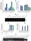
Analysis of telomere length and X-inactivation in CRES cells. (A): Relative telomere length and TERT expression in nuclear donor skin fibroblasts, CRES cell lines and in vitro fertilization (IVF)-derived ORMES-22, -23, and -7 as determined by quantitative polymerase chain reaction (q-PCR). (B): X-inactivation detected by reverse transcription-PCR analysis of XIST expression in monkey nuclear donor male and female fibroblasts, CRES cells, and ORMES-22. (C): Relative XIST expression in fibroblasts, CRES cells, and ORMES-22. (D): XIST expression levels in monkey IVF-derived XX and XY ES cell lines. OR refers to ORMES cell lines [9]. (E): XIST expression in somatic cell nuclear transfer and IVF-produced male and female monkey blastocysts. The data represents the mean ± SEM (n = 4). Abbreviations: GAPDH, glyceraldehyde 3-phosphate dehydrogenase; TERT, ribonucleoprotein complex telomerase.

References

    1. Takahashi K, Tanabe K, Ohnuki M, et al. Induction of pluripotent stem cells from adult human fibroblasts by defined factors. Cell. 2007;131:861–872. - PubMed
    1. Yu J, Vodyanik MA, Smuga-Otto K, et al. Induced pluripotent stem cell lines derived from human somatic cells. Science. 2007;318:1917–1920. - PubMed
    1. Park IH, Zhao R, West JA, et al. Reprogramming of human somatic cells to pluripotency with defined factors. Nature. 2008;451:141–146. - PubMed
    1. Huangfu D, Maehr R, Guo W, et al. Induction of pluripotent stem cells by defined factors is greatly improved by small-molecule compounds. Nat Biotechnol. 2008;26:795–797. - PMC - PubMed
    1. Byrne JA, Pedersen DA, Clepper LL, et al. Producing primate embryonic stem cells by somatic cell nuclear transfer. Nature. 2007;450:497–502. - PubMed

Publication types