

Neuroimaging in pediatric traumatic brain injury: current and future predictors of functional outcome
- PMID: 19489082
- PMCID: PMC3167090
- DOI: 10.1002/ddrr.62
Neuroimaging in pediatric traumatic brain injury: current and future predictors of functional outcome
Abstract
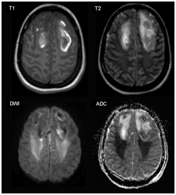
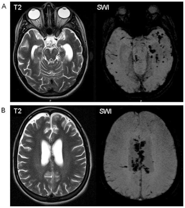
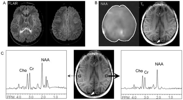
Although neuroimaging has long played a role in the acute management of pediatric traumatic brain injury (TBI), until recently, its use as a tool for understanding and predicting long-term brain-behavior relationships after TBI has been limited by the relatively poor sensitivity of routine clinical imaging for detecting diffuse axonal injury (DAI). Newer magnetic resonance-based imaging techniques demonstrate improved sensitivity to DAI. Early research suggests that these techniques hold promise for identifying imaging predictors and correlates of chronic function, both globally and within specific neuropsychological domains. In this review, we describe the principles of new, advanced imaging techniques including diffusion weighted and diffusion tensor imaging, susceptibility weighted imaging, and (1)H-magnetic resonance spectroscopy. In addition, we summarize current research demonstrating their early success in establishing relationships between imaging measures and functional outcomes after TBI. With the ongoing research, these imaging techniques may allow earlier identification of possible chronic sequelae of tissue injury for each child with TBI, thereby facilitating efficacy and efficiency in delivering successful rehabilitation services.
(c) 2009 Wiley-Liss, Inc.
Figures
Similar articles
-
Advanced neuroimaging in traumatic brain injury: an overview.Neurosurg Focus. 2019 Dec 1;47(6):E17. doi: 10.3171/2019.9.FOCUS19652. Neurosurg Focus. 2019. PMID: 32364704 Review.
-
Susceptibility weighted imaging and its relationship to outcome after pediatric traumatic brain injury.Cortex. 2013 Feb;49(2):591-8. doi: 10.1016/j.cortex.2012.08.015. Epub 2012 Sep 3. Cortex. 2013. PMID: 23062584
-
Use of advanced neuroimaging techniques in the evaluation of pediatric traumatic brain injury.Dev Neurosci. 2006;28(4-5):309-26. doi: 10.1159/000094157. Dev Neurosci. 2006. PMID: 16943654 Review.
-
Diffusion-weighted imaging improves outcome prediction in pediatric traumatic brain injury.J Neurotrauma. 2008 Oct;25(10):1153-62. doi: 10.1089/neu.2007.0494. J Neurotrauma. 2008. PMID: 18842104
-
A review of magnetic resonance imaging and diffusion tensor imaging findings in mild traumatic brain injury.Brain Imaging Behav. 2012 Jun;6(2):137-92. doi: 10.1007/s11682-012-9156-5. Brain Imaging Behav. 2012. PMID: 22438191 Free PMC article. Review.
Cited by
-
Diffusion tensor imaging of the cingulum bundle in children after traumatic brain injury.Dev Neuropsychol. 2010;35(3):333-51. doi: 10.1080/87565641003696940. Dev Neuropsychol. 2010. PMID: 20446136 Free PMC article.
-
Ability of the PILOT score to predict 6-month functional outcome in pediatric patients with moderate-severe traumatic brain injury.J Pediatr Surg. 2020 Jul;55(7):1238-1244. doi: 10.1016/j.jpedsurg.2019.06.022. Epub 2019 Jul 8. J Pediatr Surg. 2020. PMID: 31327541 Free PMC article.
-
Online problem-solving therapy for executive dysfunction after child traumatic brain injury.Pediatrics. 2013 Jul;132(1):e158-66. doi: 10.1542/peds.2012-4040. Epub 2013 Jun 10. Pediatrics. 2013. PMID: 23753094 Free PMC article. Clinical Trial.
-
Pediatric Traumatic Brain Injury: Characteristic Features, Diagnosis, and Management.Neurol Med Chir (Tokyo). 2017 Feb 15;57(2):82-93. doi: 10.2176/nmc.ra.2016-0191. Epub 2017 Jan 20. Neurol Med Chir (Tokyo). 2017. PMID: 28111406 Free PMC article. Review.
-
Pediatric head trauma: an extensive review on imaging requisites and unique imaging findings.Eur J Trauma Emerg Surg. 2018 Jun;44(3):351-368. doi: 10.1007/s00068-017-0838-y. Epub 2017 Sep 15. Eur J Trauma Emerg Surg. 2018. PMID: 28916852 Review.
References
-
- Ashikaga R, Araki Y, Ishida O. MRI of head injury using FLAIR. Neuroradiology. 1997;39:239–242. - PubMed
-
- Ashwal S, Holshouser BA, Shu SK, et al. Predictive value of proton magnetic resonance spectroscopy in pediatric closed head injury. Pediatr Neurol. 2000;23:114–125. - PubMed
-
- Babikian T, Freier MC, Tong KA, et al. Susceptbility weighted imaging: neuropsychologic outcome and pediatric head injury. Pediatr Neurol. 2005;33:184–194. - PubMed
-
- Babikian T, Freier MC, Ashwal S, et al. MR spectroscopy: predicting long-term neuropsychological outcome following pediatric TBI. J Magn Reson Imaging. 2006;24:801–811. - PubMed
